Classification of Lacrimal Punctal Stenosis and Its Related Histopathological Feature in Patients with Epiphora
- Affiliations
-
- 1Department of Ophthalmology, Han Heart Hospital, Changwon, Korea.
- 2Department of Ophthalmology, Dong-A University College of Medicine, Busan, Korea. hbahn@dau.ac.kr
- 3Department of Pathology, Dong-A University College of Medicine, Busan, Korea.
- KMID: 2390279
- DOI: http://doi.org/10.3341/kjo.2016.0129
Abstract
- PURPOSE
To evaluate the classification of punctal stenosis based on the shape of the external punctum, clinical characteristics and histopathologic features.
METHODS
Patients who experienced tearing and were diagnosed with punctal stenosis were evaluated in this study. Punctal stenosis was classified according to the shape of the lower external punctum, which included membranous type, slit type, horseshoe type, and pinpoint type. Tear meniscus height, 2% fluorescein dye disappearance test and lacrimal pathway irrigation were measured or performed. For treatment, a punctal snip operation and silicone tube placement were performed, and the peripunctal histopathological findings were evaluated.
RESULTS
Punctal stenosis was classified into four types: membranous type (17 eyes, 21.5%), slit type (11 eyes, 13.9%), horseshoe type (25 eyes, 31.6%), and pinpoint type (26 eyes, 32.9%). The tear meniscus was significantly higher, and the 2% fluorescein dye disappeared significantly more slowly in the punctal stenosis group. However, correlation of the tear meniscus height and 2% fluorescein dye disappearance test with the punctum shape was not statistically significant. A history of previous chemotherapy was significantly associated with the occurrence of punctal stenosis, especially the membranous type (p < 0.05). Histopathologic evaluation of the punctum showed differences between the punctum types. Pinpoint puncta exhibited a high density of muscle fibers, while they were faintly visible in the membranous type.
CONCLUSIONS
Acquired punctal stenosis has various shapes, and the major types of stenotic puncta exhibited unique histopathologic features. Punctal stenosis and its pathophysiology may be related to multiple factors, such as age and systemic 5-fluorouracil chemotherapy history.
MeSH Terms
Figure
-
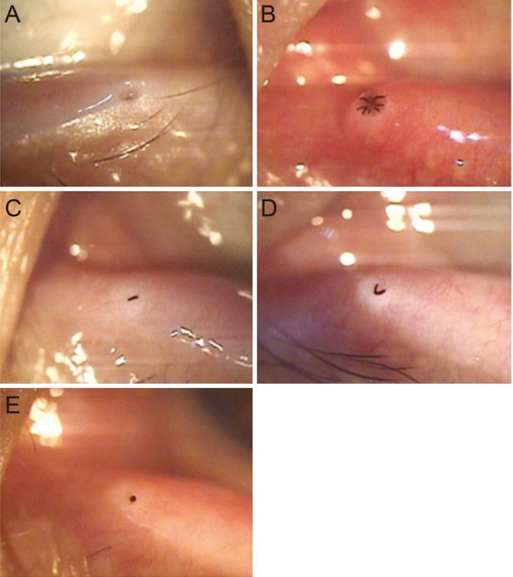
Fig. 1 The shape of a normal punctum and a stenotic or obstructed punctum. (A) Normal punctual opening, (B) membranous type punctum, (C) slit type punctum, (D) horseshoe type punctum, and (E) pinpoint type punctum (×32).
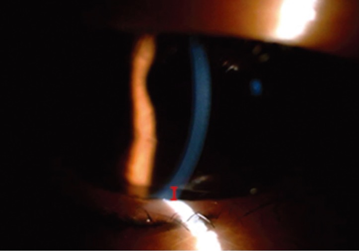
Fig. 2 Measurement of tear meniscus height with slit lamp. Tear meniscus height (vertical red bar) measured with silt lamp midsection along the lower eyelid directly below the center of the pupil (×16).
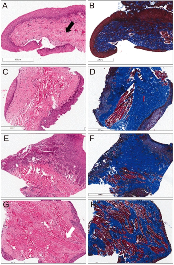
Fig. 3 Representative histopathologic findings according to classification of punctal stenosis. (A,B) Faint muscle fiber and abundant connective tissue (black arrow) of membranous type. (C,D) Moderate amount of connective tissue and muscle fibers of slit type. (E,F) Thick mucosal layer with squamous epithelial cells and a moderate amount of connective tissue with inflammation and muscle fibers of horseshoe type. (G,H) Prominent, high-density muscle fibers (white arrow) beneath mucosal layer of pinpoint type. (A,C,E,G) H&E and (B,D,F,H) Masson trichrome. Bar length = 500 µm.
Reference
-
1. Kakizaki H, Takahashi Y, Iwaki M, et al. Punctal and canalicular anatomy: implications for canalicular occlusion in severe dry eye. Am J Ophthalmol. 2012; 153:229–237. PMID: 21982102.
Article2. Lipham WJ, Tawfik HA, Dutton JJ. A histologic analysis and three-dimensional reconstruction of the muscle of Riolan. Ophthal Plast Reconstr Surg. 2002; 18:93–98.
Article3. Soiberman U, Kakizaki H, Selva D, Leibovitch I. Punctal stenosis: definition, diagnosis, and treatment. Clin Ophthalmol. 2012; 6:1011–1018. PMID: 22848141.
Article4. Kashkouli MB, Beigi B, Astbury N. Acquired external punctal stenosis: surgical management and long-term follow-up. Orbit. 2005; 24:73–78. PMID: 16191791.
Article5. Kristan RW. Treatment of lacrimal punctal stenosis with a one-snip canaliculotomy and temporary punctal plugs. Arch Ophthalmol. 1988; 106:878–879. PMID: 3390040.
Article6. Kashkouli MB, Beigi B, Murthy R, Astbury N. Acquired external punctal stenosis: etiology and associated findings. Am J Ophthalmol. 2003; 136:1079–1084. PMID: 14644218.
Article7. Bukhari A. Prevalence of punctal stenosis among ophthalmology patients. Middle East Afr J Ophthalmol. 2009; 16:85–87. PMID: 20142967.
Article8. Mainville N, Jordan DR. Etiology of tearing: a retrospective a nalysis of referrals to a tertiary care oculoplastics practice. Ophthal Plast Reconstr Surg. 2011; 27:155–157.9. Carter KD, Nelson CC, Martonyi CL. Size variation of the lacrimal punctum in adults. Ophthal Plast Reconstr Surg. 1988; 4:231–233.
Article10. Yoon KC, Jeong SK, Park YG. Study of lacrimal punctal size in normal adults. J Korean Ophthalmol Soc. 1997; 38:1916–1920.11. Kim EJ, Shin DS, Mun HJ, et al. Outcomes of anterior-side rectangular 4-snip punctoplasty for patients with punctal stenosis. J Korean Ophthalmol Soc. 2013; 54:1803–1809.
Article12. Patel S, Wallace I. Tear meniscus height, lower punctum lacrimale, and the tear lipid layer in normal aging. Optom Vis Sci. 2006; 83:731–739. PMID: 17041318.
Article13. McCulley JP, Shine WE. Changing concepts in the diagnosis and management of blepharitis. Cornea. 2000; 19:650–658. PMID: 11009317.
Article14. Shahid H, Sandhu A, Keenan T, Pearson A. Factors affecting outcome of punctoplasty surgery: a review of 205 cases. Br J Ophthalmol. 2008; 92:1689–1692. PMID: 18786958.
Article15. Hurwitz JJ. Disease of the punctum. In : Hurwitz JJ, editor. The lacrimal system. Philadelphia: Lippincott-Raven;1996. p. 149–153.16. Fezza JP, Wesley RE, Klippenstein KA. The treatment of punctal and canalicular stenosis in patients on systemic 5-FU. Ophthalmic Surg Lasers. 1999; 30:105–108. PMID: 10037204.
Article17. Brink HM, Beex LV. Punctal and canalicular stenosis associated with systemic fluorouracil therapy: report of five cases and review of the literature. Doc Ophthalmol. 1995; 90:1–6.18. McNab AA. Lacrimal canalicular obstruction associated with topical ocular medication. Aust N Z J Ophthalmol. 1998; 26:219–223. PMID: 9717753.
Article19. Edelstein J, Reiss G. The wedge punctoplasty for treatment of punctal stenosis. Ophthalmic Surg. 1992; 23:818–821. PMID: 1494436.
Article20. Caravella LP Jr, Burns JA, Zangmeister M. Punctal-canalicular stenosis related to systemic fluorouracil therapy. Arch Ophthalmol. 1981; 99:284–286. PMID: 7469866.
Article21. Prasad S, Kamath GG, Phillips RP. Lacrimal canalicular stenosis associated with systemic 5-fluorouacil therapy. Acta Ophthalmol Scand. 2000; 78:110–113. PMID: 10726804.22. Ali MJ, Mohapatra S, Mulay K, et al. Incomplete punctal canalisation: the external and internal punctal membranes. Outcomes of membranotomy and adjunctive procedures. Br J Ophthalmol. 2013; 97:92–95. PMID: 23134708.
Article23. Yuen SJ, Oley C, Sullivan TJ. Lacrimal outflow dysgenesis. Ophthalmology. 2004; 111:1782–1790. PMID: 15350337.
Article24. Lyons CJ, Rosser PM, Welham RA. The management of punctal agenesis. Ophthalmology. 1993; 100:1851–1855. PMID: 8259286.
Article25. Whitnall SE. The lacrimal apparatus. In : Whitnall SE, editor. The anatomy of the human orbit and accessory organs of vision. Oxford: Oxford University Press;1921. p. 223–252.26. Kirk RC. Developmental anomalies of the lacrimal passages; a review of the literature and presentation of three unusual cases. Am J Ophthalmol. 1956; 42:227–232.27. Port AD, Chen YT, Lelli GJ Jr. Histopathologic changes in punctal stenosis. Ophthal Plast Reconstr Surg. 2013; 29:201–204.
Article
- Full Text Links
-
- Actions
-
Cited
- CITED
-
- Close
- Share
-
- Similar articles
-
- A Simple Test for Epiphora Caused by Punctal Stenosis
- Silicone Intubation for Treatment of Punctal Stenosis
- The Surgical Results of Dacryocystorhinostomy with Internal Punctoplasty for Common Canalicular Obstruction
- The Comparison of Punctoplasty and Silicone Tube Intubation in Patients with Punctal Obstruction
- Congenital Punctal Agenesis associated with Syndaotyly in a Family
