Optimizing the reconstruction filter in cone-beam CT to improve periodontal ligament space visualization: An in vitro study
- Affiliations
-
- 1Graduate School of Medicine, Nagoya University, Japan.
- 2Department of Periodontology, School of Dentistry, Aichi Gakuin University, Japan. to-hishi@dpc.agu.ac.jp
- 3Division of Radiology, Dental Hospital, Aichi Gakuin University, Japan.
- 4Department of Oral and Maxillofacial Radiology, School of Dentistry, Aichi Gakuin University, Japan.
- KMID: 2390085
- DOI: http://doi.org/10.5624/isd.2017.47.3.199
Abstract
- PURPOSE
Evaluation of alveolar bone is important in the diagnosis of dental diseases. The periodontal ligament space is difficult to clearly depict in cone-beam computed tomography images because the reconstruction filter conditions during image processing cause image blurring, resulting in decreased spatial resolution. We examined different reconstruction filters to assess their ability to improve spatial resolution and allow for a clearer visualization of the periodontal ligament space.
MATERIALS AND METHODS
Cone-beam computed tomography projections of 2 skull phantoms were reconstructed using 6 reconstruction conditions and then compared using the Thurstone paired comparison method. Physical evaluations, including the modulation transfer function and the Wiener spectrum, as well as an assessment of space visibility, were undertaken using experimental phantoms.
RESULTS
Image reconstruction using a modified Shepp-Logan filter resulted in better sensory, physical, and quantitative evaluations. The reconstruction conditions substantially improved the spatial resolution and visualization of the periodontal ligament space. The difference in sensitivity was obtained by altering the reconstruction filter.
CONCLUSION
Modifying the characteristics of a reconstruction filter can generate significant improvement in assessments of the periodontal ligament space. A high-frequency enhancement filter improves the visualization of thin structures and will be useful when accurate assessment of the periodontal ligament space is necessary.
Keyword
MeSH Terms
Figure
-
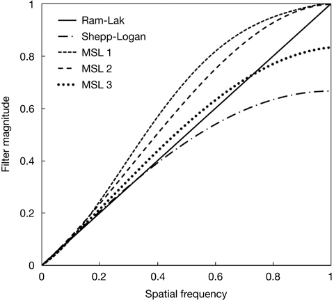
Fig. 1 Reconstruction filters prepared for observation of the periodontal ligament space. Modified Shepp-Logan (MSL) 1 shows the most highly enhanced high-frequency component, MSL 2 shows medium enhancement, and MSL 3 shows the least-enhanced high-frequency component.

Fig. 2 Diagram of the periodontal ligament phantom.

Fig. 3 Typical default images from the Alphard apparatus, and images reconstructed by each filter. A: Default, B: Ram-Lak, C: Shepp-Logan, D: modified Shepp-Logan (MSL) 1, E: MSL 2, F: MSL 3.
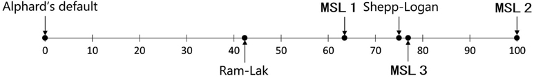
Fig. 4 The scale value calculated by the Thurstone paired-comparison method. A disparity in the scale value larger than 28 was considered a significant difference.
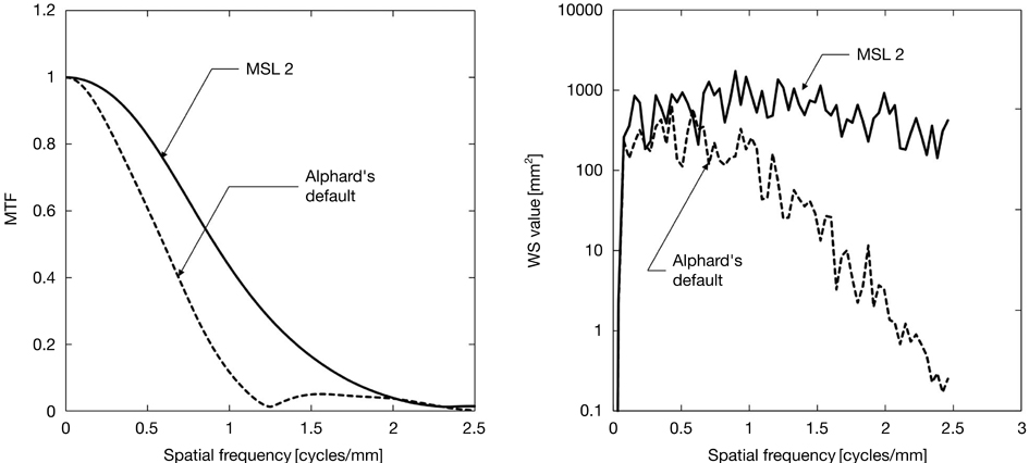
Fig. 5 The modulation transfer function (MTF) and Wiener spectrum (WS) of a modified Shepp-Logan (MSL) 2 image and the default image obtained using the Alphard apparatus.
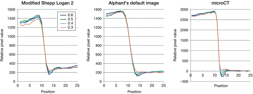
Fig. 6 Gray-value profiles of the simulated periodontal ligament space obtained by polar transformation.
Reference
-
1. Corbet EF, Ho DK, Lai SM. Radiographs in periodontal disease diagnosis and management. Aust Dent J. 2009; 54:Suppl 1. S27–S43.
Article2. Gröndhal HG, Huumonen S. Radiographic manifestations of periapical inflammatory lesions: how new radiological techniques may improve endodontic diagnosis and treatment planning. Endod Topics. 2004; 8:55–67.3. Andersson L, Blomlöf L, Lindskog S, Feiglin B, Hammarström L. Tooth ankylosis. Clinical, radiographic and histological assessments. Int J Oral Surg. 1984; 13:423–431.4. de Faria Vasconcelos K, Evangelista KM, Rodrigues CD, Estrela C, de Sousa TO, Silva MA. Detection of periodontal bone loss using cone beam CT and intraoral radiography. Dentomaxillofac Radiol. 2012; 41:64–69.
Article5. Esmaeli F, Shirmohammadi A, Faramarzie M, Abolfazli N, Rasouli H, Fallahi S. Determination of vertical interproximal bone loss topography: correlation between indirect digital radiographic measurement and clinical measurement. Iran J Radiol. 2012; 9:83–87.
Article6. Baksi BG. Measurement accuracy and perceived quality of imaging systems for the evaluation of periodontal structures. Odontology. 2008; 96:55–60.
Article7. Fuhrmann RA, Wehrbein H, Langen HJ, Diedrich PR. Assessment of the dentate alveolar process with high resolution computed tomography. Dentomaxillofac Radiol. 1995; 24:50–54.
Article8. Hishikawa T, Izumi M, Naitoh M, Yoshinari N, Kawase H, Matsuoka M, et al. Effects of the vertical projection angle in intraoral radiography on the detection of furcation involvement of the mandibular first molar. Oral Radiol. 2011; 27:102–107.
Article9. Misch KA, Yi ES, Sarment DP. Accuracy of cone beam computed tomography for periodontal defect measurements. J Periodontol. 2006; 77:1261–1266.
Article10. Walter C, Kaner D, Berndt DC, Weiger R, Zitzmann NU. Three-dimensional imaging as a pre-operative tool in decision making for furcation surgery. J Clin Periodontol. 2009; 36:250–257.
Article11. Naitoh M, Yamada S, Noguchi T, Ariji E, Nagao J, Mori K, et al. Three-dimensional display with quantitative analysis in alveolar bone resorption using cone-beam computerized tomography for dental use: a preliminary study. Int J Periodontics Restorative Dent. 2006; 26:607–612.12. Bayat S, Talaeipour AR, Sarlati F. Detection of simulated periodontal defects using cone-beam CT and digital intraoral radiography. Dentomaxillofac Radiol. 2016; 45:20160030.
Article13. Nemtoi A, Czink C, Haba D, Gahleitner A. Cone beam CT: a current overview of devices. Dentomaxillofac Radiol. 2013; 42:20120443.
Article14. Ozmeric N, Kostioutchenko I, Hägler G, Frentzen M, Jervøe-Storm PM. Cone-beam computed tomography in assessment of periodontal ligament space: in vitro study on artificial tooth model. Clin Oral Investig. 2008; 12:233–239.15. Jervøe-Storm PM, Hagner M, Neugebauer J, Ritter L, Zöller JE, Jepsen S, et al. Comparison of cone-beam computerized tomography and intraoral radiographs for determination of the periodontal ligament in a variable phantom. Oral Surg Oral Med Oral Pathol Oral Radiol Endod. 2010; 109:e95–e101.
Article16. Bushberg JT, Seibert JA, Leidholdt EM, Boone JM. The essential physics of medical imaging. 2nd ed. Philadelphia: Lippincott Williams & Wilkins;2002. p. 368–369.17. Hsieh J, Nett B, Yu Z, Sauer K, Thibault JB, Bouman CA. Recent advances in CT image reconstruction. Curr Radiol Rep. 2013; 1:39–51.
Article18. Feldkamp LA, Davis LC, Kress JW. Practical cone-beam algorithm. J Opt Soc Am A. 1984; 1:612–619.
Article19. Lee SW, Lee CL, Cho HM, Park HS, Kim DH, Choi YN, et al. Effects of reconstruction parameters on image noise and spatial resolution in cone-beam computed tomography. J Korean Phys Soc. 2011; 59:2825–2832.
Article20. Worthy S. High resolution computed tomography of the lungs. BMJ. 1995; 310:615–616.
Article21. Rezvani N, Aruliah D, Jackson K, Moseley D, Siewerdsen J. SU-FF-I-16: OSCaR: an open-source cone-beam CT reconstruction tool for imaging research. Med Phys. 2007; 34:2341.
Article22. Ramachandran GN, Lakshminarayanan AV. Three-dimensional reconstruction from radiographs and electron micrographs: application of convolutions instead of Fourier transforms. Proc Natl Acad Sci U S A. 1971; 68:2236–2240.
Article23. Shepp LA, Logan BF. The Fourier reconstruction of a head section. IEEE Trans Nucl Sci. 1974; 21:21–43.
Article24. Thurstone LL. A law of comparative judgment. Psychol Rev. 1927; 34:273–286.
Article25. Scheffe H. An analysis of variance for paired comparisons. J Am Stat Assoc. 2012; 47:381–400.
Article26. Rossmann K. Point spread-function, line spread-function, and modulation transfer function. Tools for the study of imaging systems. Radiology. 1969; 93:257–272.27. Nickoloff EL. Measurement of the PSF for a CT scanner: appropriate wire diameter and pixel size. Phys Med Biol. 1988; 33:149–155.
Article28. Boedeker KL, Cooper VN, McNitt-Gray MF. Application of the noise power spectrum in modern diagnostic MDCT: part I. Measurement of noise power spectra and noise equivalent quanta. Phys Med Biol. 2007; 52:4027–4046.
Article29. Boedeker KL, McNitt-Gray MF. Application of the noise power spectrum in modern diagnostic MDCT: part II. Noise power spectra and signal to noise. Phys Med Biol. 2007; 52:4047–4061.
Article30. Barrett J, Keat N. Artifacts in CT: recognition and avoidance. Radiographics. 2004; 24:1679–1691.
Article
- Full Text Links
-
- Actions
-
Cited
- CITED
-
- Close
- Share
-
- Similar articles
-
- Comparison of conventional imaging techniques and CBCT for periodontal evaluation: A systematic review
- Analysis of the priority of anatomic structures according to the diagnostic task in cone-beam computed tomographic images
- Commentary on "Reliability of two different presurgical preparation methods for implant dentistry based on panoramic radiography and cone-beam computed tomography in cadavers"
- Cone-beam Reconstruction using Limited EPID Projections for Seeds Localization
- Three-dimensional imaging modalities in endodontics
