Imaging Sci Dent.
2017 Sep;47(3):157-164. 10.5624/isd.2017.47.3.157.
Distances from the root apices of posterior teeth to the maxillary sinus and mandibular canal in patients with skeletal open bite: A cone-beam computed tomography study
- Affiliations
-
- 1Dental Division, Suan Phueng Hospital, Ratchaburi, Thailand.
- 2Department of Orthodontics and Pediatric Dentistry, Faculty of Dentistry, Chiang Mai University, Chiang Mai, Thailand.
- 3Department of Oral Biology and Diagnostic Sciences, Faculty of Dentistry, Chiang Mai University, Chiang Mai, Thailand. ajanhom@gmail.com
- KMID: 2390080
- DOI: http://doi.org/10.5624/isd.2017.47.3.157
Abstract
- PURPOSE
This study determined and compared the distances from the maxillary root apices of posterior teeth to the floor of the maxillary sinus, or maxillary sinus distances (MSDs), and the distances from the mandibular root apices of the posterior teeth to the mandibular canal, or mandibular canal distances (MCDs), in Thai subjects with skeletal open bite and skeletal normal bite.
MATERIALS AND METHODS
Pretreatment cone-beam computed tomography (CBCT) images were obtained from 30 Thai orthodontic patients (15 patients with skeletal normal bite and 15 with skeletal open bite) whose ages ranged from 14 to 28 years. The CBCT images of the patients were processed and measured using the Romexis Viewer program. The MSDs and MCDs from the root apices of the maxillary and mandibular second premolar, first molar, and second molar to the maxillary sinus floor or the mandibular canal were measured perpendicularly to the occlusal plane. The Student t test was used for comparisons between the 2 groups.
RESULTS
The greatest mean MSDs were from the root apex of the second premolars in both groups, whereas the least mean MSDs were from the mesiobuccal root apex of the second molars. The greatest mean MCDs were from the mesial root apex of the first molars, whereas the least mean MCDs were from the distal root apex of the second molars.
CONCLUSION
There were no differences in the mean MSDs or the mean MCDs between the skeletal normal bite group and the skeletal open bite group.
MeSH Terms
Figure
-

Fig. 1 Screen capture shows the orientation of the cone-beam computed tomography images of the right maxillary first molar for the measurements. A. Coronal view. B. Sagittal view. C. Axial view.
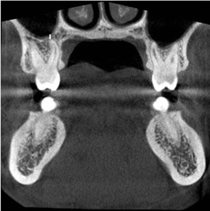
Fig. 2 Screen capture shows the distance between the root apex of the maxillary posterior teeth and maxillary sinus floor (white line).
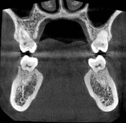
Fig. 3 Screen capture shows the distance (white line) between the root apex of the mandibular posterior teeth and mandibular canal (dotted white circle).
Reference
-
1. Fuhrmann R, Bucker A, Diedrich P. Radiological assessment of artificial bone defects in the floor of the maxillary sinus. Dentomaxillofac Radiol. 1997; 26:112–116.
Article2. Kilic C, Kamburoglu K, Yuksel SP, Ozen T. An assessment of the relationship between the maxillary sinus floor and the maxillary posterior teeth root tips using dental cone-beam computerized tomography. Eur J Dent. 2010; 4:462–467.
Article3. Sharan A, Madjar D. Correlation between maxillary sinus floor topography and related root position of posterior teeth using panoramic and cross-sectional computed tomography imaging. Oral Surg Oral Med Oral Pathol Oral Radiol Endod. 2006; 102:375–381.
Article4. Kwak HH, Park HD, Yoon HR, Kang MK, Koh KS, Kim HJ. Topographic anatomy of the inferior wall of the maxillary sinus in Koreans. Int J Oral Maxillofac Surg. 2004; 33:382–388.
Article5. de Oliveira-Santos C, Souza PH, de Azambuja Berti-Couto S, Stinkens L, Moyaert K, Rubira-Bullen IR, et al. Assessment of variations of the mandibular canal through cone beam computed tomography. Clin Oral Investig. 2012; 16:387–393.
Article6. Oliveira-Santos C, Capelozza AL, Dezzoti MS, Fischer CM, Poleti ML, Rubira-Bullen IR. Visibility of the mandibular canal on CBCT cross-sectional images. J Appl Oral Sci. 2011; 19:240–243.
Article7. Cangialosi TJ. Skeletal morphologic features of anterior open bite. Am J Orthod. 1984; 85:28–36.
Article8. Howe RB. First molar radicular bone near the maxillary sinus: a comparison of CBCT analysis and gross anatomic dissection for small bony measurement. Oral Surg Oral Med Oral Pathol Oral Radiol Endod. 2009; 108:264–269.
Article9. Kim TS, Caruso JM, Christensen H, Torabinejad M. A comparison of cone-beam computed tomography and direct measurement in the examination of the mandibular canal and adjacent structures. J Endod. 2010; 36:1191–1194.
Article10. Hassan BA. Reliability of periapical radiographs and orthopantomograms in detection of tooth root protrusion in the maxillary sinus: correlation results with cone beam computed tomography. J Oral Maxillofac Res. 2010; 1:e6.
Article11. Daimaruya T, Nagasaka H, Umemori M, Sugawara J, Mitani H. The influences of molar intrusion on the inferior alveolar neurovascular bundle and root using the skeletal anchorage system in dogs. Angle Orthod. 2001; 71:60–70.12. Jung YH, Cho BH. Assessment of the relationship between the maxillary molars and adjacent structures using cone beam computed tomography. Imaging Sci Dent. 2012; 42:219–224.
Article13. Ariji Y, Obayashi N, Goto M, Izumi M, Naitoh M, Kurita K, et al. Roots of the maxillary first and second molars in horizontal relation to the alveolar cortical plates and maxillary sinus: computed tomography assessment for infection spread. Clin Oral Investig. 2006; 10:35–41.14. Eberhardt JA, Torabinejad M, Christiansen EL. A computed tomographic study of the distances between the maxillary sinus floor and the apices of the maxillary posterior teeth. Oral Surg Oral Med Oral Pathol. 1992; 73:345–346.
Article15. Yamada T, Ishihama K, Yasuda K, Hasumi-Nakayama Y, Ito K, Yamaoka M, et al. Inferior alveolar nerve canal and branches detected with dental cone beam computed tomography in lower third molar region. J Oral Maxillofac Surg. 2011; 69:1278–1282.
Article16. Kovisto T, Ahmad M, Bowles W. Proximity of the mandibular canal to the tooth apex. J Endod. 2011; 37:311–315.
Article17. Sato I, Ueno R, Kawai T, Yosue T. Rare courses of the mandibular canal in the molar regions of the human mandible: a cadaveric study. Okajimas Folia Anat Jpn. 2005; 82:95–102.
Article
- Full Text Links
-
- Actions
-
Cited
- CITED
-
- Close
- Share
-
- Similar articles
-
- Relationship of the maxillary posterior teeth and maxillary sinus floor in different skeletal growth patterns: A cone-beam computed tomographic study of 1600 roots
- Reliability of panoramic radiography in predicting proximity of third molars to the mandibular canal: A comparison using cone-beam computed tomography
- Cone-beam computed tomography for the assessment of root–crown ratios of the maxillary and mandibular incisors in a Korean population
- Root surface areas of maxillary permanent teeth in anterior normal overbite and anterior open bite assessed using cone-beam computed tomography
- Assessment of the relationship between the maxillary molars and adjacent structures using cone beam computed tomography
