Plexiform Angiomyxoid Myofibroblastic Tumor of the Stomach: a Rare Case
- Affiliations
-
- 1Department of Surgery, Samsung Medical Center, Sungkyunkwan University School of Medicine, Seoul, Korea. jmoon.bae@samsung.com
- 2Department of Pathology, Samsung Medical Center, Sungkyunkwan University School of Medicine, Seoul, Korea.
- KMID: 2389832
- DOI: http://doi.org/10.5230/jgc.2017.17.e22
Abstract
- Plexiform angiomyxoid myofibroblastic tumor (PAMT) of the stomach is a very rare mesenchymal tumor of the gastrointestinal tract. We report a case of asymptomatic gastric PAMT that was pathologically confirmed after surgical resection. The tumor had a multinodular plexiform growth pattern, bland-looking spindle cells, and an Alcian blue-positive myxoid stromal matrix rich in small blood vessels. Immunohistochemistry analysis revealed that the tumor cells of the PAMT were positive for smooth muscle actin (SMA) and negative for c-kit, CD34, S-100 protein, epithelial membrane antigen (EMA), and desmin. PAMT should be differentiated from other submucosal tumors of the stomach by immunohistochemical findings. Considering the benign features of this tumor, observation without resection may be an option for the treatment of PAMT if the tumor is asymptomatic.
Keyword
MeSH Terms
Figure
-
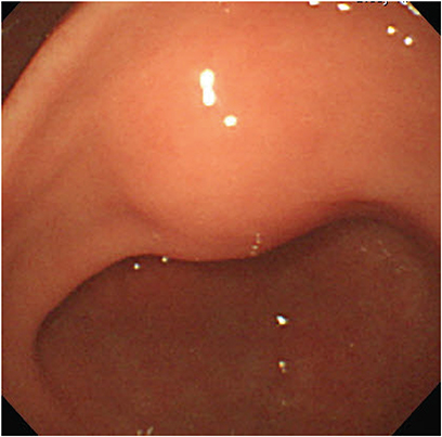
Fig. 1 Esophagogastroduodenoscopy shows a smooth, normal mucosa-covered elevated lesion at the gastric antrum.

Fig. 2 Computed tomography shows a well-marginated cystic lesion, suspected to be a GIST, glomus tumor, or neuroendocrine tumor. GIST = gastrointestinal stromal tumor.
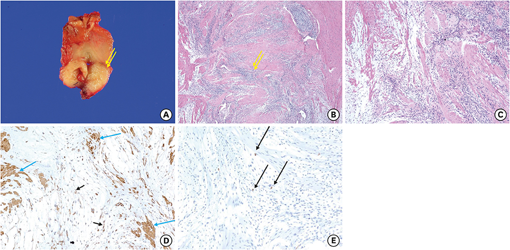
Fig. 3 (A) Tumor surgically removed by laparoscopic wedge resection. Cut section shows a well-defined whitish and firm mass. (B) Tumor demonstrates infiltrative growth of cellular areas into smooth muscle bundles in the muscularis propria. (C) Tumor shows loose myxoid areas (left part) and cellular areas (right part) admixed with smooth muscle cells. (D) Smooth muscle fibers and tumor cells positive for smooth muscle actin (×200). (E) Tumor cells negative for c-kit. Mast cells were positively stained as an internal positive control (arrows).
Reference
-
1. Miettinen M, Makhlouf HR, Sobin LH, Lasota J. Plexiform fibromyxoma: a distinctive benign gastric antral neoplasm not to be confused with a myxoid GIST. Am J Surg Pathol. 2009; 33:1624–1632.2. Takahashi Y, Shimizu S, Ishida T, Aita K, Toida S, Fukusato T, et al. Plexiform angiomyxoid myofibroblastic tumor of the stomach. Am J Surg Pathol. 2007; 31:724–728.3. Kim A, Bae YK, Shin HC, Choi JH. Plexiform angiomyxoid myofibroblastic tumor of the stomach: a case report. J Korean Med Sci. 2011; 26:1508–1511.4. Kang Y, Jung W, Do IG, Lee EJ, Lee MH, Kim KM, et al. Plexiform angiomyxoid myofibroblastic tumor of the stomach: report of two cases and review of the literature. Korean J Pathol. 2012; 46:292–296.5. Yoshida A, Klimstra DS, Antonescu CR. Plexiform angiomyxoid tumor of the stomach. Am J Surg Pathol. 2008; 32:1910–1912.6. Quero G, Musarra T, Carrato A, Fici M, Martini M, Dei Tos AP, et al. Unusual focal keratin expression in plexiform angiomyxoid myofibroblastic tumor: a case report and review of the literature. Medicine (Baltimore). 2016; 95:e4207.7. Banerjee N, Gupta S, Dash S, Ghosh S. Plexiform angiomyxoid myofibroblastic tumour of the duodenum: a rare entity. BMJ Case Rep. 2015; 2015:bcr2015210004.8. Lee PW, Yau DT, Lau PP, Chan JK. Plexiform fibromyxoma (plexiform angiomyxoid myofibroblastic tumor) of stomach: an unusual presentation as a fistulating abscess. Int J Surg Pathol. 2014; 22:286–290.9. Vander Noot MR 3rd, Eloubeidi MA, Chen VK, Eltoum I, Jhala D, Jhala N, et al. Diagnosis of gastrointestinal tract lesions by endoscopic ultrasound-guided fine-needle aspiration biopsy. Cancer. 2004; 102:157–163.10. Schulz T, Drgac J, Chmelar C, Höhler T, Agaimy A, Vieth M. Plexiform angiomyxoid myofibroblastic tumour of the stomach. Pathologe. 2012; 33:65–69.
- Full Text Links
-
- Actions
-
Cited
- CITED
-
- Close
- Share
-
- Similar articles
-
- Plexiform Angiomyxoid Myofibroblastic Tumor of the Stomach: A Case Report
- Plexiform Angiomyxoid Myofibroblastic Tumor of the Stomach: Report of a Case and Review of the Literature
- Plexiform Angiomyxoid Myofibroblastic Tumor of the Stomach: Report of Two Cases and Review of the Literature
- Inflammatory Myofibroblastic Tumor of Nasal Septum after Septoplasty: A Case Report
- A Case of Myxoid Plexiform Fibrohistiocytic Tumor
