Sonographic Findings of Polyneuropathy Associated With Cerebrotendinous Xanthomatosis: A Case Report
- Affiliations
-
- 1Department of Rehabilitation Medicine, College of Medicine, The Catholic University of Korea, Seoul, Korea. minukkim@nate.com
- KMID: 2389491
- DOI: http://doi.org/10.5535/arm.2017.41.2.313
Abstract
- Cerebrotendinous xanthomatosis is a rare autosomal recessive disease that involves multiple organs, including the peripheral nervous system. The present study is the first to report the ultrasonographic findings of peripheral nerves in a patient with cerebrotendinous xanthomatosis. The patient presented with bilateral Achilles tendon enlargement and foot hypesthesia. Sonographic examination revealed hypoechoic, swollen peripheral nerves with enlarged bilateral Achilles tendons. Since the ultrasonographic findings revealed peripheral involvement, the diagnosis of cerebrotendinous xanthomatosis was established after laboratory and genetic studies along with clinical findings.
MeSH Terms
Figure
-
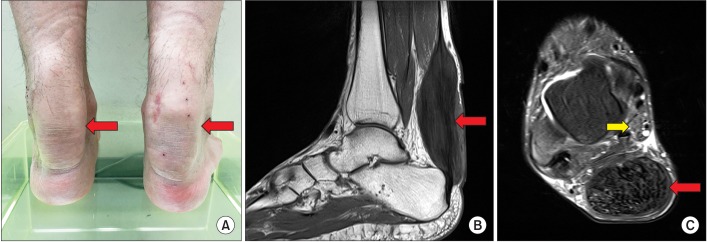
Fig. 1 Clinical photograph demonstrated bilateral fusiform enlargement of the Achilles tendon (red arrow) (A). A sagittal T1-weighted image (B) and an axial T2-weighted image of the ankle magnetic resonance imaging (C) showed a diffuse, hypointense mass occupying the entire Achilles tendon (red arrow) with a swollen tibial nerve (yellow arrow) behind the medial malleolus. The xanthoma measured 13.8 cm in length, 3.2 cm in anteroposterior diameter, and 4.4 cm in the transverse dimension.

Fig. 2 Schematic illustrations show sonographic image of the patient (A, D, G). Transverse ultrasonography scan of the peripheral nerves in the patient (B, E, H) and in a normal subject whose anthropometry was similar to that of the patient (C, F, I). In the patient, the tibial nerve (arrow) behind the medial malleolus was swollen, compared with that in the normal subject (A–C). The median nerve (arrow) at the wrist and elbow was also swollen, compared with that in the normal control (D–I). MM, medial malleolus; TP, tibialis posterior; FDL, flexor digitorum longus; A, posterior tibial artery; V, posterior tibial vein; SCA, scaphoid bone; PIS, pisiform bone; FCR, flexor carpi radialis; FPL, flexor pollicis longus; S, flexor digitorum superficialis; D, flexor digitorum profundus; UA, ulnar artery; BA, brachial artery; B, brachialis; PT, pronator teres; U, ulna bone.
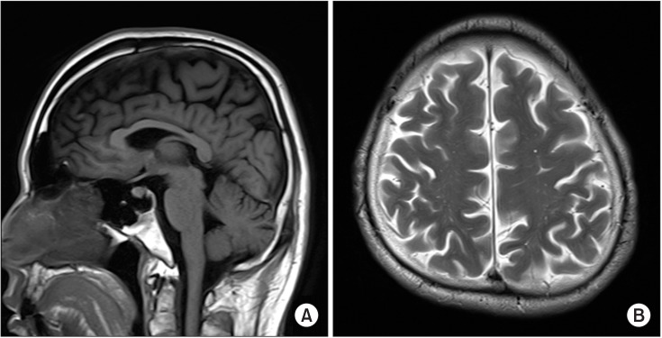
Fig. 3 On magnetic resonance imaging of the brain, the sagittal T1-weighted image showed mild shrinkage of the cerebellum (A) and the axial T2-weighted image showed slightly high signal intensity in the bilateral fronto-parieto-occipital periventricular white matter (B).
Reference
-
1. Nie S, Chen G, Cao X, Zhang Y. Cerebrotendinous xanthomatosis: a comprehensive review of pathogenesis, clinical manifestations, diagnosis, and management. Orphanet J Rare Dis. 2014; 9:179. PMID: 25424010.
Article2. Moghadasian MH. Cerebrotendinous xanthomatosis: clinical course, genotypes and metabolic backgrounds. Clin Invest Med. 2004; 27:42–50. PMID: 15061585.3. Wang Z, Yuan Y, Zhang W, Zhang Y, Feng L. Cerebrotendinous xanthomatosis with a compound heterozygote mutation and severe polyneuropathy. Neuropathology. 2007; 27:62–66. PMID: 17319284.
Article4. Pilo B, de Blas G, Sobrido MJ, Navarro C, Grandas F, Barrero FJ, et al. Neurophysiological study in cerebrotendinous xanthomatosis. Muscle Nerve. 2011; 43:531–536. PMID: 21404287.
Article5. Ginanneschi F, Mignarri A, Mondelli M, Gallus GN, Del Puppo M, Giorgi S, et al. Polyneuropathy in cerebrotendinous xanthomatosis and response to treatment with chenodeoxycholic acid. J Neurol. 2013; 260:268–274. PMID: 22878431.
Article6. Verrips A, van Engelen BG, ter Laak H, Gabreels-Festen A, Janssen A, Zwarts M, et al. Cerebrotendinous xanthomatosis: controversies about nerve and muscle: observations in ten patients. Neuromuscul Disord. 2000; 10:407–414. PMID: 10899446.7. Ohnishi A, Yamashita Y, Goto I, Kuroiwa Y, Murakami S, Ikeda M. De- and remyelination and onion bulb in cerebrotendinous xanthomatosis. Acta Neuropathol. 1979; 45:43–45. PMID: 760364.
Article8. Qrimli M, Ebadi H, Breiner A, Siddiqui H, Alabdali M, Abraham A, et al. Reference values for ultrasonograpy of peripheral nerves. Muscle Nerve. 2016; 53:538–544. PMID: 26316047.
Article
