Ann Rehabil Med.
2017 Apr;41(2):257-265. 10.5535/arm.2017.41.2.257.
Changes in Resting Calcaneal Stance Position Angle Following Insole Fitting in Children With Flexible Flatfoot
- Affiliations
-
- 1Department of Physical and Rehabilitation Medicine, Inha University School of Medicine, Incheon, Korea. rmkmo@inha.ac.kr
- KMID: 2389484
- DOI: http://doi.org/10.5535/arm.2017.41.2.257
Abstract
OBJECTIVE
To clarify the relationship of the initial radiologic and a biomechanical parameter at first clinical visit, and define the effectiveness of modified insole, following insole fitting in children with flexible flatfoot.
METHODS
Children aged less than 13 years with flexible flatfoot were enrolled. The total number of subjects was 66 (33 boys, 33 girls). The subjects were divided into 5 subgroups, based on age: 1-2, 3-4, 5-6, 7-9, and 10-12 years. The mean time period between the initial & final examination for their resting calcaneal stance position angle (RCSPA) was 24 months. Radiography quantified the deformity by measuring angles, including the talometatarsal angle, the metatarsal angle, and the calcaneal pitch angle.
RESULTS
From the angles measured on radiographs, only the talometatarsal angle showed a statistically significant correlation to the initial RCSPA (r=-0.578 for right side, r=-0.524 for left side; p<0.01). The mean RCSPA improved in all subgroups of subjects following insole fitting. Moreover, in children younger than 7 years, the improvement in RCSPA from the insole fitting was greater compared to children aged 7 years and older.
CONCLUSION
The insole has additionally beneficial effects in all populations younger than 13 years. However, there might exist a hidden effect of normal structural pedal alignment during growth accompanied with bony maturation and developmental process. To date, it is controversial whether the treatment of flexible flatfoot is necessary in the vast majority of cases, or simple observation and advice to parents would suffice.
Keyword
Figure
-
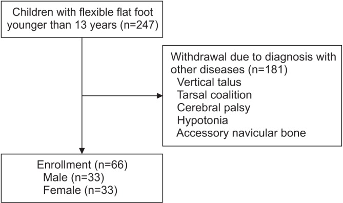
Fig. 1 Flow chart of study selection.
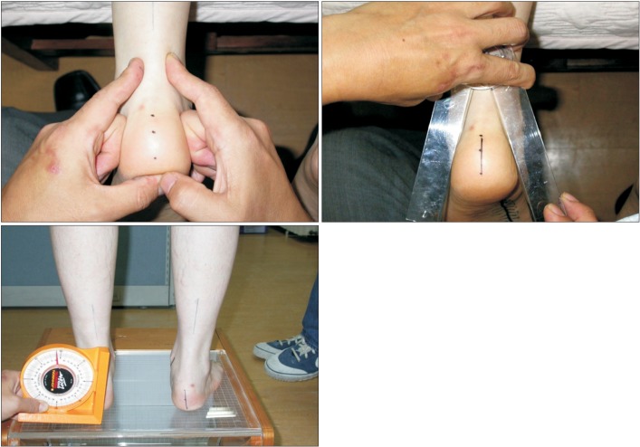
Fig. 2 Measurement of the resting calcaneal stance position angle (RCSPA).
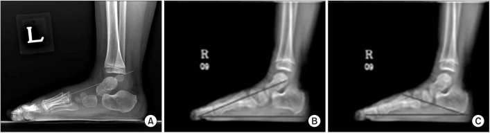
Fig. 3 (A) Talometatarsal angle, (B) metatarsal angle, and (C) calcaneal pitch angle.
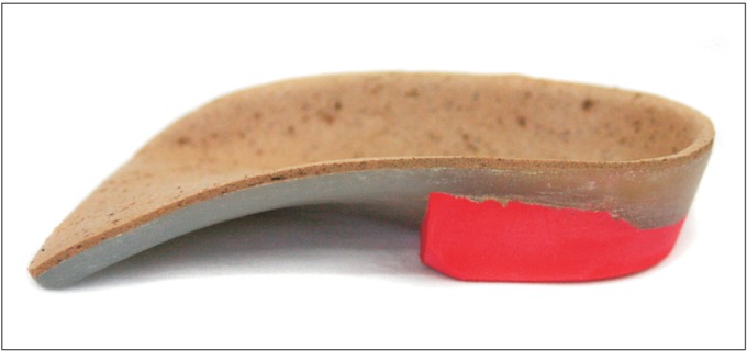
Fig. 4 Custom-made rigid foot orthosis manufactured on the basis of the inverted technique (Biomechanics, Goyang, Korea).
Cited by 1 articles
-
Effect of Foot Orthoses in Children With Symptomatic Flexible Flatfoot Based on Ultrasonography of the Ankle Invertor and Evertor Muscles
Dong Joon Cho, So Young Ahn, Soo-Kyung Bok
Ann Rehabil Med. 2021;45(6):459-470. doi: 10.5535/arm.21137.
Reference
-
1. Lee JH, Sung IY, Yoo JY. Clinical or radiologic measurements and 3-D gait analysis in children with pes planus. Pediatr Int. 2009; 51:201–205. PMID: 19405916.
Article2. Chen YC, Lou SZ, Huang CY, Su FC. Effects of foot orthoses on gait patterns of flat feet patients. Clin Biomech (Bristol, Avon). 2010; 25:265–270.
Article3. Kanatli U, Yetkin H, Cila E. Footprint and radiographic analysis of the feet. J Pediatr Orthop. 2001; 21:225–228. PMID: 11242255.
Article4. Sobel E, Levitz SJ, Caselli MA, Tran M, Lepore F, Lilja E, et al. Reevaluation of the relaxed calcaneal stance position: reliability and normal values in children and adults. J Am Podiatr Med Assoc. 1999; 89:258–264. PMID: 10349290.
Article5. Halabchi F, Mazaheri R, Mirshahi M, Abbasian L. Pediatric flexible flatfoot: clinical aspects and algorithmic approach. Iran J Pediatr. 2013; 23:247–260. PMID: 23795246.6. Mosca VS. Flexible flatfoot in children and adolescents. J Child Orthop. 2010; 4:107–121. PMID: 21455468.
Article7. Harris EJ, Vanore JV, Thomas JL, Kravitz SR, Mendelson SA, Mendicino RW, et al. Diagnosis and treatment of pediatric flatfoot. J Foot Ankle Surg. 2004; 43:341–373. PMID: 15605048.
Article8. Cappello T, Song KM. Determining treatment of flat-feet in children. Curr Opin Pediatr. 1998; 10:77–81. PMID: 9529644.
Article9. Sullivan JA. Pediatric flatfoot: evaluation and management. J Am Acad Orthop Surg. 1999; 7:44–53. PMID: 9916191.
Article10. Tang SF, Chen CH, Wu CK, Hong WH, Chen KJ, Chen CK. The effects of total contact insole with forefoot medial posting on rearfoot movement and foot pressure distributions in patients with flexible flatfoot. Clin Neurol Neurosurg. 2015; 129(Suppl 1):S8–S11. PMID: 25683316.
Article11. Aboutorabi A, Saeedi H, Kamali M, Farahmand B, Eshraghi A, Dolagh RS. Immediate effect of orthopedic shoe and functional foot orthosis on center of pressure displacement and gait parameters in juvenile flexible flat foot. Prosthet Orthot Int. 2014; 38:218–223. PMID: 23986466.
Article12. Banwell HA, Mackintosh S, Thewlis D. Foot orthoses for adults with flexible pes planus: a systematic review. J Foot Ankle Res. 2014; 7:23. PMID: 24708560.
Article13. Jay RM, Schoenhaus HD, Seymour C, Gamble S. The Dynamic Stabilizing Innersole System (DSIS): the management of hyperpronation in children. J Foot Ankle Surg. 1995; 34:124–131. PMID: 7599609.
Article14. Mereday C, Dolan CM, Lusskin R. Evaluation of the University of California Biomechanics Laboratory shoe insert in “flexible” pes planus. Clin Orthop Relat Res. 1972; 82:45–58. PMID: 5011037.
Article15. Selby-Silverstein L, Hillstrom HJ, Palisano RJ. The effect of foot orthoses on standing foot posture and gait of young children with Down syndrome. NeuroRehabilitation. 2001; 16:183–193. PMID: 11790903.
Article16. Shin JB, Kim SW, You S, Lee SK, Kim HS. Biomechanic analysis of lower extremities in children and teenagers with pes planus. J Korean Acad Rehabil Med. 2008; 32:154–159.17. Ko YJ, Kim HW. Physical examination of foot and ankle. J Korean Assoc Pain Med. 2006; 5:1–7.18. Gould N, Moreland M, Alvarez R, Trevino S, Fenwick J. Development of the child's arch. Foot Ankle. 1989; 9:241–245. PMID: 2731836.
Article19. Staheli LT, Chew DE, Corbett M. The longitudinal arch: a survey of eight hundred and eighty-two feet in normal children and adults. J Bone Joint Surg Am. 1987; 69:426–428. PMID: 3818704.20. Evans AM, Rome K. A Cochrane review of the evidence for non-surgical interventions for flexible pediatric flat feet. Eur J Phys Rehabil Med. 2011; 47:69–89. PMID: 21448121.21. Kim SB, Yoon K, Park HS, Kwak H, Ha NJ, Park JS. Radiologic measurement of flatfoot. J Korean Acad Rehabil Med. 2000; 24:995–1001.22. Fabry G. Clinical practice: static, axial, and rotational deformities of the lower extremities in children. Eur J Pediatr. 2010; 169:529–534. PMID: 20052491.23. Yagerman SE, Cross MB, Positano R, Doyle SM. Evaluation and treatment of symptomatic pes planus. Curr Opin Pediatr. 2011; 23:60–67. PMID: 21169838.24. Labovitz JM. The algorithmic approach to pediatric flexible pes planovalgus. Clin Podiatr Med Surg. 2006; 23:57–76. PMID: 16598910.
Article25. Pfeiffer M, Kotz R, Ledl T, Hauser G, Sluga M. Prevalence of flat foot in preschool-aged children. Pediatrics. 2006; 118:634–639. PMID: 16882817.
Article26. Kwon JY, Myerson MS. Management of the flexible flat foot in the child: a focus on the use of osteotomies for correction. Foot Ankle Clin. 2010; 15:309–322. PMID: 20534358.
Article27. Mortazavi SJ, Espandar R, Baghdadi T. Flatfoot in children: how to approach? Iran J Pediatr. 2007; 17:163–170.
- Full Text Links
-
- Actions
-
Cited
- CITED
-
- Close
- Share
-
- Similar articles
-
- Effects of Biomechanical Foot Orthoses on the Resting Calcaneal Stance Position Angle in Flatfoot Patients
- Effect of Foot Orthoses in Children With Symptomatic Flexible Flatfoot Based on Ultrasonography of the Ankle Invertor and Evertor Muscles
- Effect of Custom-Molded Foot Orthoses on Foot Pain and Balance in Children With Symptomatic Flexible Flat Feet
- Effect of Pressure Based Customized 3-Dimensional Printing Insole in Pediatric Flexible Flat Foot Patients
- Adult Idiopathic Flexible Flat Foot Treated with Medial Sliding Calcaneal Osteotomy and Subtalar Arthroereisis: Report of 1 Case
