Ann Dermatol.
2017 Oct;29(5):661-663. 10.5021/ad.2017.29.5.661.
A Case of Acquired Mucinous Nevus in Nevus Lipomatosus Cutaneous Superficialis
- Affiliations
-
- 1Department of Dermatology, Hallym University Sacred Heart Hospital, Anyang, Korea. dermakkh@naver.com
- KMID: 2388932
- DOI: http://doi.org/10.5021/ad.2017.29.5.661
Abstract
- No abstract available.
Figure
-

Fig. 1 (A) Localized various sized yellowish papuloplaques on the buttock. (B) Solitary well defined 0.3×0.3 cm pedunculated skin colored papule on the buttock.
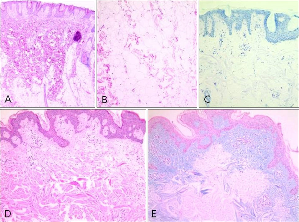
Fig. 2 From plaque lesion. (A, B) Hyperkeratosis and acanthosis of the epidermis, and clusters of adipocytes among collagen fibers (H&E; A: ×10, B: ×100). (C) Negative staining with toluidine blue (×100). From papule lesion. (D) Hyperkeratosis, mild acanthosis, and mucin deposition in papillary dermis (H&E, ×100). (E) Positive staining with alcian blue at pH 2.5 in papillary dermis (×100).
Reference
-
1. Hoffmann E, Zurhelle E. Über einen naevus lipomatosus cutaneus superficialis der linken glutaalgegend. Arch Dermatol Syphilol. 1921; 130:327–333.
Article2. Perez-Crespo M, Lopez-Navarro N, Betlloch I, Herrera E, Niveiro M, Gallego E. Acquired and familial mucinous nevus. Int J Dermatol. 2011; 50:1283–1285.
Article3. Redondo Bellón P, Vázquez-Doval J, Idoate M, Quintanilla E. Mucinous nevus. J Am Acad Dermatol. 1993; 28:797–798.
Article4. Song BH, Park S, Park EJ, Kwon IH, Kim KH, Kim KJ. Mucinous nevus with fat: an unusual case report and literature review. Am J Dermatopathol. 2012; 34:e146–e148.5. Carapeto FJ, Chárlez L, Marrón J, Grasa MP, Marrón SE. Infantile and progressive papular mucinosis. Med Cutan Ibero Lat Am. 1985; 13:525–530.
- Full Text Links
-
- Actions
-
Cited
- CITED
-
- Close
- Share
-
- Similar articles
-
- Nevus Lipomatosus Superficialis on the Left Leg
- A Case of Nevus Lipomatosus Cutaneous Superficialis
- Familial Case of Nevus Lipomatosus Superficialis
- Nipple Shaped Solitary Nevus Lipomatosus Cutaneous Superficialis: Report of a Case and a Review of the Korean Literatures
- Nevus Lipomatosus Cutaneous Superficialis: Report of A Case
