A Case of Recurrent Myxomatous Corneal Degeneration Treated by Interferon Alpha-2b
- Affiliations
-
- 1Department of Ophthalmology, Chonbuk National University Medical School, Jeonju, Korea. you2ic@daum.net
- 2Research Institute of Clinical Medicine, Chonbuk National University, Jeonju, Korea.
- 3Biomedical Research Institute, Chonbuk National University Hospital, Jeonju, Korea.
- KMID: 2388191
- DOI: http://doi.org/10.3341/jkos.2017.58.8.998
Abstract
- PURPOSE
To report a case of recurrent myxomatous corneal degeneration after pterygectomy.
CASE SUMMARY
A 65-year-old man with a history of abdominal keloid was referred to our hospital for assessment of a well-circumscribed, gelatinous, whitish corneal mass on the nasal corneal area of the left eye that appeared one month prior. The patient had undergone pterygectomy on his left eye 2 years ago. The patient experienced mild foreign body sensation. The other anterior segment and fundus examination of the left eye were both normal. We diagnosed the case as keloid and we performed excisional biopsy of the corneal mass. Histologic findings revealed proliferation of myxoid-appearing material in the anterior corneal stroma. On immunohistochemical examination, sections were stained positive for actin and calretinin, and negative for S-100. We diagnosed the tumor as myxomatous corneal degeneration. After six months, a recurrent mass was found on the previously excised site. Re-excisional biopsy and topical interferon α-2b treatment were then performed in response. After re-excision, there was no recurrence or complications during one year follow-up.
CONCLUSIONS
Myxomatous corneal degeneration should be considered during differential diagnosis of an elevated, whitish, gelatinous lesion of the cornea with previous history of trauma or operation, such as pterygectomy. An interferon α-2b topical treatment is useful for recurrent corneal myomatous degeneration.
MeSH Terms
Figure
-

Figure 1 Slit-lamp findings of corneal mass from the left eye of a 65-year-old man. (A) At the first visit, well circumscribed whitish, movable, and partially transparent corneal mass on the nasal corneal area. (B) Mass was removed well on the cornea by excision.
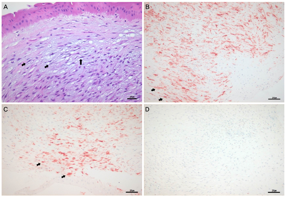
Figure 2 Immunohistochemical staining on permanent paraffin slides of corneal mass. (A) Spindle cell proliferation with reticular fiber in prominent hypovascular myxoid stroma (H&E, ×400) (arrows), Scale bar, 20 µm. (B) Positive for actin stain, ×200 (arrows), Scale bar, 20 µm. (C) Positive for calretinin stain, ×200 (arrows), Scale bar, 20 µm. (D) Negative for S-100 stain, ×200, Scale bar, 20 µm.
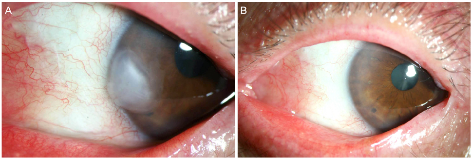
Figure 3 Anterior segment photography of recurrence 6 months after excision. (A) Recurrent corneal mass on the same site. (B) Note the clear cornea without recurrence or complication at 12 months after re-excision.
Reference
-
1. Bulkley BH, Hutchins GM. Atrial myxomas: a fifty year review. Am Heart J. 1979; 97:639–643.2. Park SJ, Lee MJ, Sung MS, et al. A case of conjunctival myxoma. J Korean Ophthalmol Soc. 2008; 49:1676–1679.3. Baek JW, Jung SK, Paik JS, Yang SW. A case of conjunctival myxoma invading the caruncle. J Korean Ophthalmol Soc. 2013; 54:954–957.4. Horie Y, Ikawa S, Okamoto I, et al. Myxoma of the conjunctiva: a case report and a review of the literature. Jpn J Ophthalmol. 1995; 39:77–82.5. Chang HJ. Superficial corneal growth. JAMA. 2011; 305:2467–2468.6. Lim KS, Wee SW, Kim JC. Treatment of an 8-mm myxoma using acellular corneal tissue. Korean J Ophthalmol. 2014; 28:86–90.7. Hansen LH, Prause JU, Ehlers N, Heegaard S. Primary corneal myxoma. Acta Ophthalmol Scand. 2004; 82:224–227.8. Lee BT, Lee JH, Kim SH, Cho MY. One case of corneal myxoma. J Korean Ophthalmol Soc. 1994; 35:202–205.9. Ireland DC, Soule EH, Ivins JC. Myxoma of somatic soft tissues. A report of 58 patients, 3 with multiple tumors and fibrous dysplasia of bone. Mayo Clin Proc. 1973; 48:401–410.10. Terracciano LM, Mhawech P, Suess K, et al. Calretinin as a marker for cardiac myxoma. Diagnostic and histogenetic considerations. Am J Clin Pathol. 2000; 114:754–759.11. Belliveau MJ, Liao WN, Brownstein S, et al. Myxomatous corneal degeneration: a clinicopathological study of six cases and a review of the literature. Surv Ophthalmol. 2012; 57:264–271.12. Wollensak G, Green WR, Seiler T. Corneal myxoma. Jpn J Ophthalmol. 2002; 46:193–197.13. Lo GG, Biswas J, Rao NA, Font RL. Corneal myxoma. Case report and review of the literature. Cornea. 1990; 9:174–178.14. Chawla B, Agarwal A, Kashyap S, Tandon R. Diagnosis and management of corneal keloid. Clin Exp Ophthalmol. 2007; 35:855–857.15. Shields CL, Shields JA. Tumors of the conjunctiva and cornea. Surv Ophthalmol. 2004; 49:3–24.16. Karp CL, Moore JK, Rosa RH Jr. Treatment of conjunctival and corneal intraepithelial neoplasia with topical interferon alpha-2b. Ophthalmology. 2001; 108:1093–1098.
- Full Text Links
-
- Actions
-
Cited
- CITED
-
- Close
- Share
-
- Similar articles
-
- Effect of alpha-Interferon 2b on Chronic Hepatitis B Patients with High Serum ALT
- Bell's Palsy during Interferon Therapy for Chronic Hapatitis C Infection
- Generalized Eczema-like Eruption in a Patient Treated with Interferon Alpha-2b and Ribavirin for Chronic Hepatitis C
- Development of thyroid autoimmunity after administration of recombinant human interferon alpha 2b in patients with chronic viral hepatitis
- Acute Pancreatitis Associated with Pegylated Interferon alpha-2b and Ribavirin Treatment of Chronic Hepatitis C
