Managements of ankylosed incisor occurred during adolescence using alveolar bone distraction osteogenesis and decoronation: case report
- Affiliations
-
- 1Dental Clinic Center, Pusan National University Hospital, Busan, Republic of Korea. youngyng@hanmail.net
- 2Department of Orthodontics, School of Dentistry, Pusan National University, Yangsan, Republic of Korea.
- KMID: 2388021
- DOI: http://doi.org/10.14368/jdras.2017.33.2.143
Abstract
- One of the common complications of dental injury is tooth ankylosis. Unlike adults, when tooth ankylosis occurs in the adolescents, ankylosis interfered the growth of the adjacent alveolar bone, resulting in the developmental failure of the alveolar bone and subsequent open bite. The most common treatment option for ankylosed tooth is extraction. However, when prognosis of ankylosed tooth after extraction is expected to be poor due to severity of infrapositioning or prosthetic replacement cannot be performed immediately, various treatment options should be considered. This report suggests multidisciplinary treatment that might bring functionally and esthetically favorable result included alveolar bone distraction osteogenesis and decoronation of ankylosed maxillary anterior tooth with orthodontic and prosthetic treatments.
MeSH Terms
Figure
-
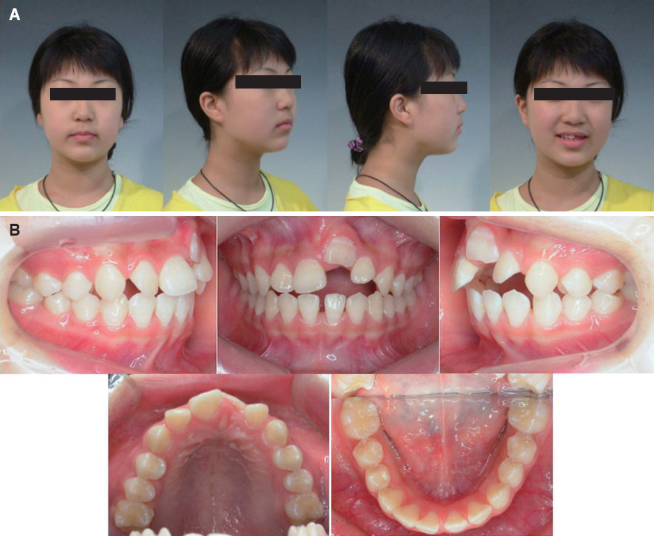
Fig. 1 (A) Pretreatment extraoral and intraoral photographs (1st treatment). The patient was 11 years old. (B) #21 was ankylosed and located infraocclusionly more than 8 mm due to trauma at 7 years. Anterior open bite was observed.

Fig. 2 (A) Pretreatment lateral cephalograph, periapical and panoramic radiographs (1st treatment). (B) Lamina dura of #21 was disappeared in radiographic. (A - C) #21 was located infraocclusionly
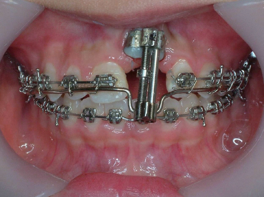
Fig. 3 Intraoral appliance for #21 distraction osteogenesis after single tooth osteotomy. Distraction was carried out 0.5 - 1.0 mm per day.
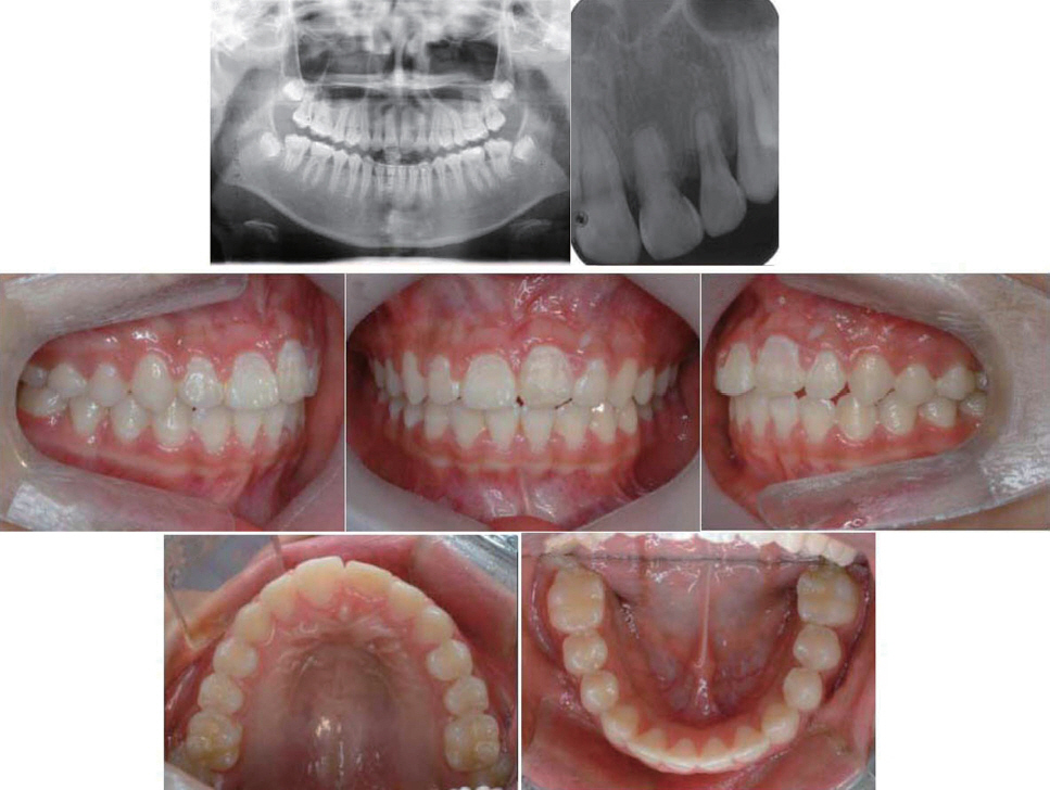
Fig. 4 Posttreatment intraoral photographs, panoramic and periapical radiographs (1st treatment).
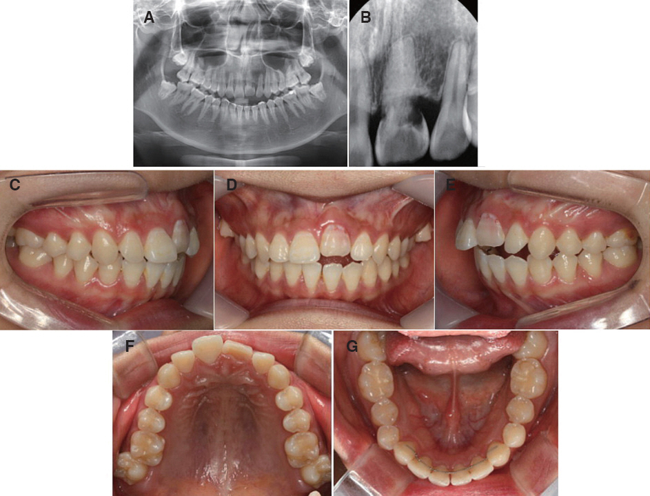
Fig. 5 (A, B) Follow-up (after 6 years) photographs, panoramic and periapical radiographs. The external root resorption on #21 was occurred. (C - G) The vertical position of #21 was infraocclusion and pink spot of cervical area was observed.

Fig. 6 Intraoral photographs during orthodontic treatment (2nd treatment).
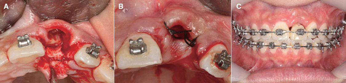
Fig. 7 Procedure of decoronation. (A, B) In order to create new marginal bone, the coronal part of the root surface was removed 2 mm below the marginal bone (C). The crown part of #21 was adjusted and placed like an artificial tooth.
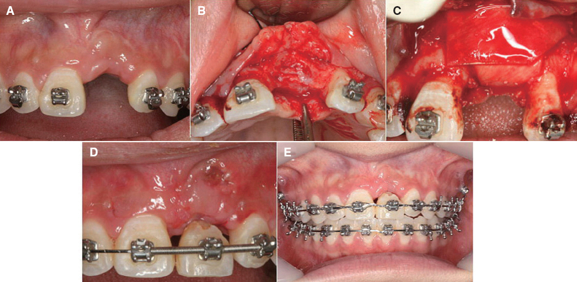
Fig. 8 Extraction of the root rest and GBR procedure. (A, B) Remaining buccal alveolar bone thickness was too thin. (C) Guided bone regeneration procedure was performed on buccal area with bovine bone material and collagen membrane. (D, E) The crown part of #21 was used like an artificial tooth and adjusted to alveolar crest contour.
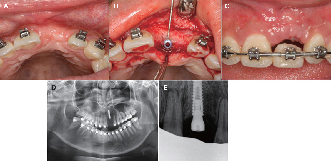
Fig. 9 After 6 months of GBR operation, implantation of fixture. (A, B) Procedure of implant placement. (C) The crown part of #21 was adjusted to alveolar crest contour. (D, E) Post-operative panoramic radiograph and standard periapical radiograph.
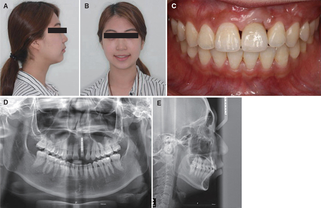
Fig. 10 (A - C) Posttreatment extraoral and intraoral photographs. The final prosthetic restoration of #21 was performed. (D, E) Posttreatment panoramic radiograph and lateral cephalograph.
Reference
-
References
1. Andersson L, Bodin I, Sörensen S. Progression of root resorption following replantation of human teeth after extended extraoral storage. Endod Dent Traumatol. 1989; 5:38–47. DOI: 10.1111/j.1600-9657.1989.tb00335.x. PMID: 2598883.2. Borssén E, Källestål C, Holm AK. Treatment time of traumatic dental injuries in a cohort of 16-yearolds in northern Sweden. Acta Odontol Scand. 2002; 60:265–70. DOI: 10.1080/00016350260248229.3. Loriato LB, Machado AW, Souki BQ, Pereira TJ. Late diagnosis of dentoalveolar ankylosis:impact on effectiveness and efficiency of orthodontic treatment. Am J Orthod Dentofacial Orthop. 2009; 135:799–808. DOI: 10.1016/j.ajodo.2007.04.040. PMID: 19524841.4. Ebeleseder KA, Friehs S, Ruda C, Pertl C, Glockner K, Hulla H. A study of replanted permanent teeth in different age groups. Endod Dent Traumatol. 1998; 14:274–8. DOI: 10.1111/j.1600-9657.1998.tb00852.x. PMID: 9972160.5. Kinzinger GS, Jänicke S, Riediger D, Diedrich PR. Orthodontic fine adjustment after vertical callus distraction of an ankylosed incisor using the floating bone concept. Am J Orthod Dentofacial Orthop. 2003; 124:582–90. DOI: 10.1016/S0889-5406(03)00569-9.6. Kofod T, Würtz V, Melsen B. Treatment of an ankylosed central incisor by single tooth dentoosseous osteotomy and a simple distraction device. Am J Orthod Dentofacial Orthop. 2005; 127:72–80. DOI: 10.1016/j.ajodo.2003.12.020. PMID: 15643418.7. Samchukov ML, Cope JB, Cherkashin AM. Craniofacial distraction osteogenesis. Mosby;2001. p. 379458.8. Gound T, O’Neal RB, del Rio CE, Levin MP. Submergence of roots for alveolar bone preservation. II. Reimplanted endodontically treated roots. Oral Surg Oral Med Oral Pathol. 1978; 46:114–122. DOI: 10.1016/0030-4220(78)90445-0.9. Johnson DL, Kelly JF, Flinton RJ, Cornell MT. Histologic evaluation of vital root retention. J Oral Surg. 1974; 32:829–33.10. Andreasen JO, Paulsen HU, Yu Z, Schwartz O. A long-term study of 370 autotransplanted premolars. Part III. Periodontal healing subsequent to transplantation. Eur J Orthod. 1990; 12:25–37. DOI: 10.1093/ejo/12.1.25. PMID: 2318260.11. Ilizarov GA. Clinical application of the tensionstress effect for limb lengthening. Clin Orthop Relat Res. 1990; (250):8–26.12. Riolo ML, Moyers RE, McNamara JA Jr, Hunter WS. An atlas of craniofacial growth:cephalometric standards from The University of Michigan School Growth Study. Ann Arbor; The University of Michigan. 1974; 101–216.13. Malmgren B, Malmgren O, Andreasen JO. Alveolar bone development after decoronation of ankylosed teeth. Endod Topics. 2006; 14:35–40. DOI: 10.1111/j.1601-1546.2008.00225.x.14. Filippi A, Pohl Y, von Arx T. Decoronation of an ankylosed tooth for preservation of alveolar bone prior to implant placement. Dent Traumatol. 2001; 17:93–5. DOI: 10.1034/j.1600-9657.2001.017002093.x. PMID: 11475952.15. Sapir S, Kalter A, Sapir MR. Decoronation of an ankylosed permanent incisor:alveolar ridge preservation and rehabilitation by an implant supported porcelain crown. Dent Traumatol. 2009; 25:346–9. DOI: 10.1111/j.1600-9657.2009.00788.x. PMID: 19583582 .
- Full Text Links
-
- Actions
-
Cited
- CITED
-
- Close
- Share
-
- Similar articles
-
- Orthodontic treatment of an ankylosed tooth; application of single tooth osteotomy and alveolar bone distraction osteogenesis
- Solutions and Prevention of Problems Arising from Alveolar Distraction Osteogenesis: 4 Case Reports
- Evaluation of augmented alveolar bone with vertical alveolar distraction osteogenesis and implant installation
- Vertical Distraction Of Alveolar Bone For Placement Of Dental Implant
- Distraction osteogenesis in patients with hemifacial microsomia
