Sonography of the Rotator Cuff: Comparison of Arm Positions
- Affiliations
-
- 1Hanmaeum Orthopaedic Clinic, Daejeon, Korea. borameos@hanmail.net
- 2Department of Orthopedic Surgery, Seoul Sacred Heart General Hospital, Seoul, Korea.
- 3Department of Orthopedic Surgery, Chungnam National University Hospital, Daejeon, Korea.
- KMID: 2387792
- DOI: http://doi.org/10.4055/jkoa.2017.52.4.336
Abstract
- PURPOSE
To evaluate the objective difference of the shoulder position during ultrasound examination regarding diagnostic value for shoulder lesion, view range and visibility.
MATERIALS AND METHODS
A prospective study was performed enrolling 312 patients who underwent diagnostic ultrasonography due to shoulder pain between January 2016 and June 2016. Examination was performed by a single orthopaedic surgeon with 5 years of musculoskeletal ultrasonography experience. Images of the longitudinal and transverse plane of the supraspinatus tendon and the nearby soft tissues (subscapularis and biceps long head tendon, subdeltoid bursa, etc.) were obtained in the three different positions, shoulder extension, modified Crass, and Crass position. The correlation between the demographic data (age, sex and body mass index) and the visual analogue scale (VAS) of the affected shoulder & the capable shoulder position was analyzed. Another orthopaedic independently measured the size of the tear and using classified the image visibility of the supraspinatus, subscapularis, and biceps long head tendon on the short-axis view from the rotator interval into I to III and X.
RESULTS
Of the 312 patients, 126 were excluded and total of 186 cases were included in this study. None of the demographic data were related to the possible arm position. However, VAS for pain was the only factor related with the number of possible arm positions during sonography. Kappa agreements for the diagnosis were mostly high of over 0.90. Grades of the short-axis view from the rotator interval in each position were mostly grade II or grade III, which refers to that the anterior portion of supraspinatus tendon, which is the most fragile portion to the tear and it was well-defined regardless of the arm position. The average longitudinal tear sizes were 1.48, 1.52, and 1.61 cm in the shoulder extension, modified Crass (Middleton), and Crass position, respectively.
CONCLUSION
Shoulder extension position during ultrasonography examination of shoulder shows similar diagnosis rate of supraspinatus tendon tear or calcific tendinitis compared to modified Crass (Middleton) or Crass position, the two well-known standard positions. It is also a useful position for patients who suffer with severe shoulder pain.
Keyword
MeSH Terms
Figure
-

Figure 1 This figure shows positioning of the arm during sonography. (A) Shoulder extension position. (B) Modified Crass position. (C) Crass position.
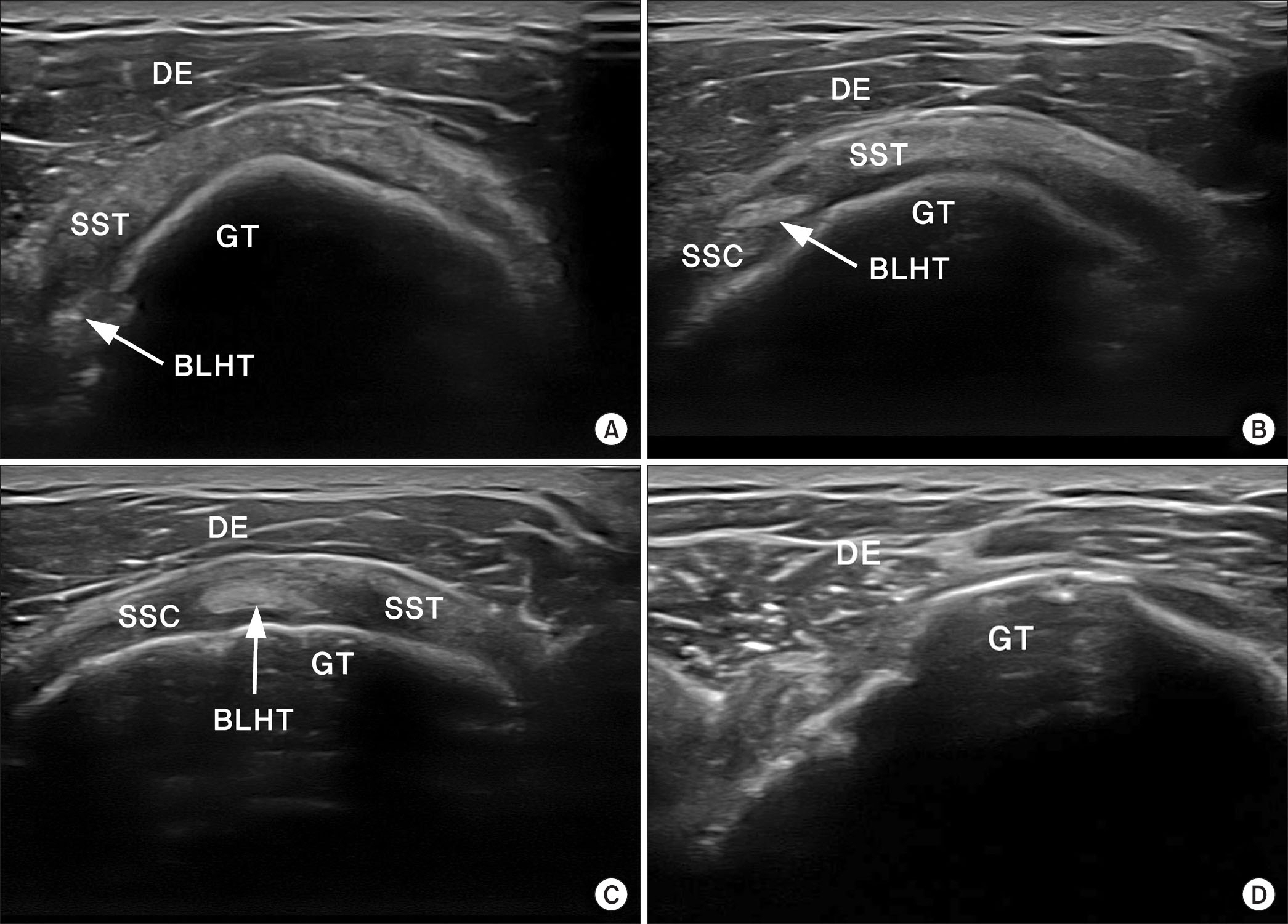
Figure 2 These sonographic images show the grading of the short-axis view of the rotator interval. (A) Grade I, when the border between the BLHT and the SST tendon is not observed prominently. (B) Grade II, when the border between the BLHT and the SST tendon is observed prominently, while the border between the BLHT and the SSC tendon is not observed prominently. (C) Grade III, when the border between the SSC tendon, the BLHT, and the SST tendon is prominently observed in one field of view. (D) Grade X, when the border is ambiguous due to a complete rupture of the SST tendon or BLHT or an extensive rupture of the rotator cuff. DE, deltoid muscle; SST, supraspinatus; GT, greater tubercle; BLHT, biceps long head tendon; SSC, subscapularis.
Reference
-
1. Bianchi S, Martinoli C. Ultrasound of the musculoskeletal system. Berlin: Springer;. 2007; 189–91.
Article2. Adler RS, Sofka CM. Percutaneous ultrasound-guided injections in the musculoskeletal system. Ultrasound Q. 2003; 19:3–12.
Article3. Cardinal E, Chhem RK, Beauregard CG. Ultrasound-guided interventional procedures in the musculoskeletal system. Radiol Clin North Am. 1998; 36:597–604.
Article4. Crass JR, Craig EV, Feinberg SB. The hyperextended internal rotation view in rotator cuff ultrasonography. J Clin Ultrasound. 1987; 15:416–20.
Article5. Ferri M, Finlay K, Popowich T, Stamp G, Schuringa P, Friedman L. Sonography of full-thickness supraspinatus tears: comparison of patient positioning technique with surgical correlation. AJR Am J Roentgenol. 2005; 184:180–4.
Article6. Singh JP. Shoulder ultrasound: what you need to know. Indian J Radiol Imaging. 2012; 22:284–92.
Article7. Shah NP, Miller TT, Stock H, Adler RS. Sonography of supraspinatus tendon abnormalities in the neutral versus Crass and modified Crass positions: a prospective study. J Ultrasound Med. 2012; 31:1203–8.8. Mack LA, Matsen FA 3rd, Kilcoyne RF, Davies PK, Sickler ME. US evaluation of the rotator cuff. Radiology. 1985; 157:205–9.
Article9. Crass JR, Craig EV, Feinberg SB. Ultrasonography of rotator cuff tears: a review of 500 diagnostic studies. J Clin Ultrasound. 1988; 16:313–27.
Article10. Wiener SN, Seitz WH Jr. Sonography of the shoulder in patients with tears of the rotator cuff: accuracy and value for selecting surgical options. AJR Am J Roentgenol. 1993; 160:103–7.
Article11. Middleton WD. Ultrasonography of the shoulder. Radiol Clin North Am. 1992; 30:927–40.12. Yablon CM, Bedi A, Morag Y, Jacobson JA. Ultrasonography of the shoulder with arthroscopic correlation. Clin Sports Med. 2013; 32:391–408.
Article13. Crass JR, Craig EV, Thompson RC, Feinberg SB. Ultrasonography of the rotator cuff: surgical correlation. J Clin Ultrasound. 1984; 12:487–91.
Article14. Middleton WD, Edelstein G, Reinus WR, Melson GL, Totty WG, Murphy WA. Sonographic detection of rotator cuff tears. Am J Roentgenol. 1985; 144:349–53.
Article15. Teefey SA, Hasan SA, Middleton WD, Patel M, Wright RW, Yamaguchi K. Ultrasonography of the rotator cuff. A comparison of ultrasonographic and arthroscopic findings in one hundred consecutive cases. J Bone Joint Surg Am. 2000; 82:498–504.16. Moon SH. A trip to the musculoskeletal ultrasound with doctor Moon. 2015. 1st ed. Seoul: Youngchang Publishing Company;p. 221–32.
- Full Text Links
-
- Actions
-
Cited
- CITED
-
- Close
- Share
-
- Similar articles
-
- Biological Characteristics of Rotator Cuff Tendon
- Revisional Rotator Cuff Repair
- Change to Type and Size of Rotator Cuff Tear Following Arthro-3D Sonography
- Intramuscular Cyst of the Rotator Cuff Associated with Tear of the Rotator Cuff: A Case Report
- Delaminated Rotator Cuff Tear: Concurrent Concept and Treatment
