Nat Prod Sci.
2017 Jun;23(2):125-131. 10.20307/nps.2017.23.2.125.
Anthocyanins from Clitoria ternatea Attenuate Food-Borne Penicillium expansum and its Potential Application as Food Biopreservative
- Affiliations
-
- 1Universiti Kuala Lumpur, Malaysian Institute of Chemical and Bioengineering Technology, Lot 1988 Kawasan Perindustrian Bandar Vendor, Taboh Naning, 78000 Alor Gajah, Melaka. wytong@unikl.edu.my
- 2Universiti Tunku Abdul Rahman, Jalan Universiti, Bandar Barat, 31900 Kampar, Perak, Malaysia.
- 3School of Distance Education, Universiti Sains Malaysia, 11800 Minden, Penang, Malaysia.
- KMID: 2387063
- DOI: http://doi.org/10.20307/nps.2017.23.2.125
Abstract
- Clitoria ternatea or Commonly known blue pea, is a perennial climber crop native to Asian countries. The current study was aimed to evaluate the antimicrobial activity C. ternatea extract on food borne microorganisms and its antifungal effect on Penicillium expansum. The extract showed significant antimicrobial activity against 3 Gram positive bacteria, 2 Gram negative bacteria and 1 filamentous fungus on disc diffusion assay. The extract also showed good biocidal effect on all Gram positive bacteria tested and P. expansum. However, the kill curve analysis revealed that the fungicidal activity of the extract against P. expansum conidia was depend on the concentration of the extract and the time of exposure of the conidia to the extract. The scanning electron micrograph of the extract treated P. expansum culture showed alterations in the morphology of fungal hyphae. The germination of P. expansum conidia was completely inhibited and conidial development was totally suppressed by the extract, suggesting the possible mode of action of anthocyanin. Besides, the extract also exhibited 5.0-log suppression of microbial growth relative to control in the rice model. The results indicate the potential use of the C. ternatea anthocyanin as food biopreservative.
MeSH Terms
Figure
-
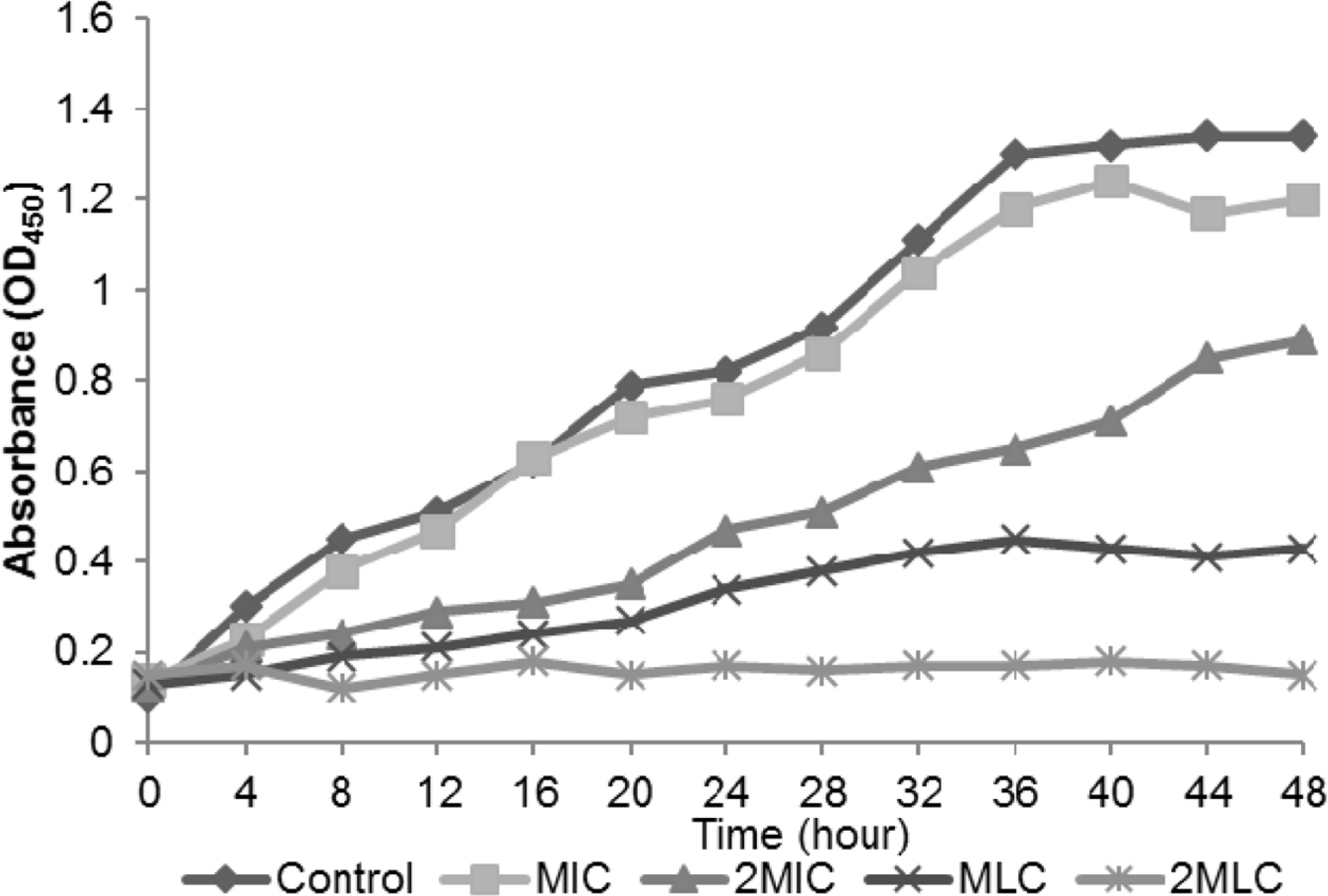
Fig. 1. The kill curve of C. ternatea extract on P. expansum. The fungicidal activity of the extract was concentration-dependent.

Fig. 2. SEM micrograph of food borne P. expansum treated with (A) sterile distilled water (B) C. ternatea extract (5000× magnification). The results suggest the irreversible damage on the conidiophore of P. expansum.
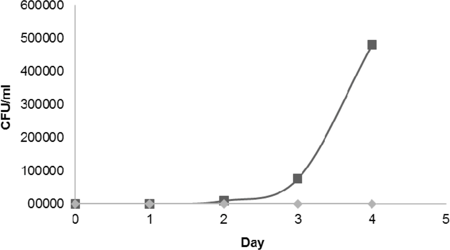
Fig. 3. The microbial load of the food model obtained throughout the 5 days of incubation period. Test substance: (◆) C. ternatea extract, (■) Distilled water control. The extract significantly inhibits the microbial growth in the rice model.
Reference
-
References
(1). Lima A. A. N. D.., Sobringho J. L. S.., Lyra M. A. M. D.., Santos F. L. A. D.., Figueiredo C. B. M.., Neto P. J. R.Int. J. Pharm. Pharm. Sci. 2015. 7:371–375.(2). Kagan C. R.ACS Nano. 2016. 10:2985–2986.(3). Kawai T.., Sekizuka T.., Yahata Y.., Kuroda M.., Kumeda Y.., Iijima Y.., Kamata Y.., Sugita-Konishi Y.., Ohnishi T.Clin. Infect. Dis. 2012. 54:1046–1052.(4). Abdul-Mutalib N. A.., Nordin S. A.., Osman M.., Ishida N.., Tashiro K.., Sakai K.., Tashiro Y.., Maeda T.., Shirai Y.Int. J. Food Microbiol. 2015. 200:57–65.(5). Spadaro D.., Lorè A.., Garibaldi A.., Gullino M. L.Postharvest Biol. Technol. 2013. 75:1–8.(6). Yu C.., Zhou T.., Sheng K.., Zeng L.., Ye C.., Yu T.., Zheng X.Int. J. Food Microbiol. 2013. 164:155–160.(7). Bhatia M.., Chahal J.., Gupta S.Int. J. Pharm. Sci. Res. 2014. 5:600–606.(8). Kungsuwan K.., Singh K.., Phetkao S.., Utama-ang N.Food Appl. Biosci. J. 2014. 2:31–46.(9). Chong F. C.., Gwee X. F.Nat. Prod. Res. 2015. 29:1485–1487.(10). Priya F. J.., Vimala J. R.., Bama R. S.., Lavanya M.World J. Pharm. Pharm. Sci. 2016. 5:753–765.(11). Tong W. Y.., Ang S. N.., Darah I.., Latiffah Z.World J. Pharm. Pharm. Sci. 2014. 3:121–132.(12). Darah I.., Chong C. L.., Tong W. Y.., Latiffah Z.., Lim S.Trop. J. Pharm. Res. 2015. 14:2091–2097.(13). Dalla Lana D. F.., Donato R. K.., Bündchen C.., Guez C. M.., Bergamo V. Z.., de Oliveira L. F.., Machado M. M.., Schrekker H. S.., Fuentefria A. M. J.Appl. Microbiol. 2015. 119:377–388.(14). Biswas B.., Rogers K.., McLaughlin F.., Daniels D.., Yadav A.Int. J. Microbiol. 2013. 2013:1–7.(15). Kamilla L.., Mnsor S. M.., Ramanathan S.., Sasidharan S.Pharm. Online. 2009. 1:731–738.(16). Nair V.., Bang W. Y.., Schreckinger E.., Andarwulan N.., Cisneros-Zevallos L. J.Agric. Food Chem. 2015. 63:6355–6365.(17). Kondo T.., Ueda M.., Goto T.Tetrahedron. 1990. 46:4749–4756.(18). Rao V. S.., Santos F. A.., Sobreira T. T.., Souza M. F.., Melo C. L.., Silveira E. R.Planta Med. 1997. 63:146–149.(19). Nikaido H.Microbiol. Mol. Biol. Rev. 2003. 67:593–656.(20). Palazzo I. C. V.., Araujo M. L. C.., Darini A. L. C. J.Clin. Microbiol. 2005. 43:179–185.(21). Kamilla L.., Mansor S. M.., Ramanathan S.., Sasidharan S.Microsc. Microanal. 2009. 15:366–372.(22). Moussa S. H.., Tayel A. A.., Al-Hassan A. A.., Farouk A. J.Mycol. 2013. 2013:753692.(23). Rafiee E.., Khodayari M.., Shahebrahimi S.., Joshaghani M. J.Mol. Catal. A. 2011. 351:204–209.
- Full Text Links
-
- Actions
-
Cited
- CITED
-
- Close
- Share
-
- Similar articles
-
- Six Species of Penicillium Associated with Blue Mold of Grape
- The Ethanolic Extract of Clitoria ternatea L. Flower Attenuates SMADs and REGs Regulation in Dyslipidemia and Diabetes Mellitus Rat Model
- Diversity of Aspergillus, Penicillium, and Talaromyces Species Isolated from Freshwater Environments in Korea
- Food-Borne Parasitic Diseases
- Prevalence and Transmission of Seed-Borne Fungi of Maize Grown in a Farm of Korea
