Ultrasonographic Findings of Pilar Sheath Acanthoma: A Case Report
- Affiliations
-
- 1Department of Radiology, Inje University College of Medicine, Busan Paik Hospital, Busan, Korea. sunjulee98@naver.com
- 2Center for Fishermen's Safety & Health, Inje University College of Medicine, Busan Paik Hospital, Busan, Korea.
- 3Department of Radiology, Hongcheon Asan Hospital, Hongcheon, Korea.
- KMID: 2386751
- DOI: http://doi.org/10.3348/jksr.2017.77.2.129
Abstract
- Pilar sheath acanthoma is a rare benign follicular hamartoma that presents with a central sinus containing keratinous material and is lined by epithelium. It typically occurs on the face, especially on the upper lip and forehead. In our case, the ultrasound (US) feature of pilar sheath acanthoma revealed a well-defined, oval hypoechoic nodule with hypoechoic capping within the dermis over the medial aspect of the calf. To the best of our knowledge, despite many reports on the clinicopathological aspects of pilar sheath acanthoma, this entity has not been well described in the radiologic literature, and US findings have not been documented. We report the US findings of a case of pilar sheath acanthoma on the calf.
Figure
-
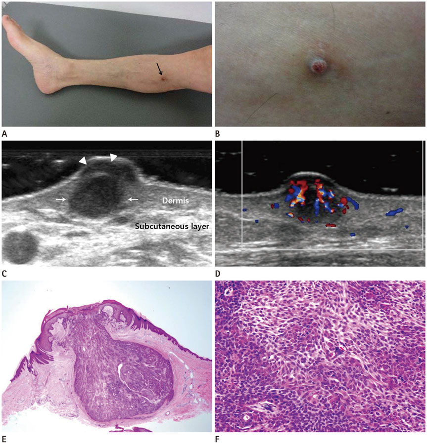
Fig. 1 Pilar sheath acanthoma on the right calf in a 56-year-old man. A, B. Photograph of the leg. Medial (A) and close-up (B) views of the papule on the right calf. A solitary, erythematous to hyperpigmented papule with a central punctum (arrow in A) is seen on the medial aspect of the right leg. C. Gray scale ultrasonography shows a 0.65 × 0.35 cm, well-defined oval hypoechoic nodule (arrows) with hypoechoic capping (arrowheads) within the dermis over the medial aspect of the right calf. D. A color Doppler image shows internal hypervascularity within the nodule. E. A photomicrograph (hematoxylin-eosin stain, × 10 ) shows a lobular patulous tumor that was connected to the epithelium and the dermis. F. A photomicrograph (hematoxylin-eosin stain, × 200) shows tumor cells consisting of mixed blue-gray (infundibular) and pink (isthmic) corneocytes. The findings are compatible with pilar sheath acanthoma.
Reference
-
1. Mehregan AH, Brownstein MH. Pilar sheath acanthoma. Arch Dermatol. 1978; 114:1495–1497.2. Lee JW, Lee HI, Kim B, Kim MN, Song KY. A case of pilar sheath acanthomas on the both cheeks. Korean J Dermatol. 2009; 47:1077–1079.3. Ackerman AB, de Viragh PA, Chongchitnant N. Neoplasms with follicular differentiation. Philadelphia: Lea & Febiger;1993. p. 509–529.4. Kushner JA, Thomas RS, Young RJ. An unusual location of a pilar sheath acanthoma. Int J Trichology. 2014; 6:185–186.5. Choi YS, Park SH, Bang D. Pilar sheath acanthoma--report of a case with review of the literature. Yonsei Med J. 1989; 30:392–395.6. Shih-Tsung CJ, Lee YY. Clinical and histological features of dilated pore: report of six cases and review of literature. Dermatol Sinica. 1993; 11:121–128.7. Beaman FD, Kransdorf MJ, Andrews TR, Murphey MD, Arcara LK, Keeling JH. Superficial soft-tissue masses: analysis, diagnosis, and differential considerations. Radiographics. 2007; 27:509–523.8. Lee HS, Joo KB, Song HT, Kim YS, Park DW, Park CK, et al. Relationship between sonographic and pathologic findings in epidermal inclusion cysts. J Clin Ultrasound. 2001; 29:374–383.9. Hwang JY, Lee SW, Lee SM. The common ultrasonographic features of pilomatricoma. J Ultrasound Med. 2005; 24:1397–1402.10. Lim HW, Im SA, Lim GY, Park HJ, Lee H, Sung MS, et al. Pilomatricomas in children: imaging characteristics with pathologic correlation. Pediatr Radiol. 2007; 37:549–555.
