Amniotic constriction band: a report of two cases with unique clinical presentations
- Affiliations
-
- 1Richardsons Dental and Craniofacial Hospital, Nagercoil, India. sunilrichardson145@gmail.com
- 2Department of National Reference, Center for Rare Craniomaxillofacial Malformations, Centre Hospitalier Régional Universitaire de Lille, Lille, France.
- KMID: 2386362
- DOI: http://doi.org/10.5125/jkaoms.2017.43.3.171
Abstract
- Amniotic constriction band is a rare clinical entity with varied manifestations that range from a combination of congenital malformations to isolated malformations that are unique to each patient. The etiology of this entity remains unknown. Herein, we highlight two cases of amniotic constriction band that presented to our unit with unique clinical characteristics. To the best of our knowledge, an isolated circumferential band of scarring on the face with ocular involvement, as demonstrated by the first case, and a combination of bilateral complete cleft lip and palate with bilateral microphthalmia, auto-amputation of the right thumb, and a constriction band on the left thumb, as demonstrated by the second case, are extremely rare presentations of amniotic constriction band that were not previously reported in the literature and therefore necessitate a special mention. We discuss potential etiologies for these cases and review the existing literature on this entity.
Keyword
Figure
-

Fig. 1 Clinical presentation of the circumferential constriction band on the face in Case 1. A. Frontal view showing the constriction band on the face. B. Right lateral view showing the extent of the constriction band on the scalp. C. Left lateral view showing the extent of the constriction band. D. Close-up view to appreciate the ocular involvement.
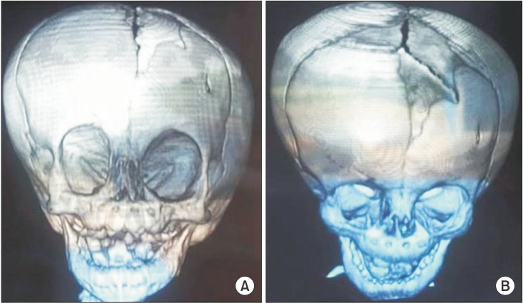
Fig. 2 Three-dimensional computed tomographic view of the facial skeleton in Case 1. A. View demonstrating the smaller dimensions of the right orbit as compared to the left. The right orbit measured 2.9×2.1 cm in greatest dimensions whereas the left orbit measured 3.0×2.6 mm in greatest dimensions. B. View demonstrating a defect in the frontal bone on the left side.
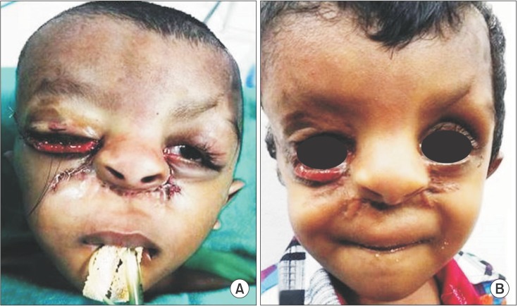
Fig. 3 Intraoperative and postoperative photographs of Case 1. A. Intraoperative photographs of bilateral oculoplastic surgery. B. Postoperative photographs at 6 months from surgery.

Fig. 4 Clinical presentation of Case 2. A. Frontal view showing bilateral complete cleft lip, premaxillary excess and bilateral microphthalmia. B. Photograph of the right hand showing auto-amputation of the thumb. C. Photograph of the left hand showing constriction band on the left thumb.

Fig. 5 Intraoperative and postoperative photographs of bilateral cleft lip repair of Case 2. A. Intraoperative view of repair using Mulliken technique. B. Postoperative view at 6 months.
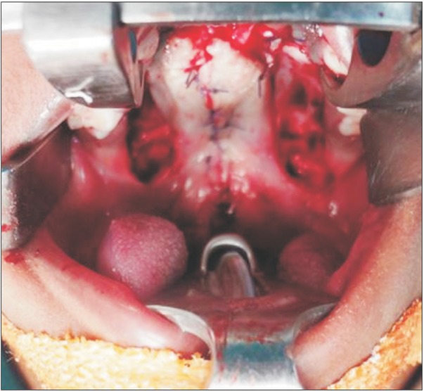
Fig. 6 Intraoperative photograph of cleft palate repair in Case 2.

Fig. 7 Intraoperative and postoperative photographs of vomerine osteotomy of Case 2. A. Intraoperative view following osteotomy and fixation. B. Postoperative view.
Reference
-
1. Ciloglu NS, Gumus N. A rare form of congenital amniotic band syndrome: total circular abdominal constriction band. Arch Plast Surg. 2014; 41:290–291. PMID: 24883282.
Article2. Matic A, Komazec J. Amniotic band syndrome. Acta Medica Medianae. 2009; 48:44–48.3. Das D, Das G, Gayen S, Konar A. Median facial cleft in amniotic band syndrome. Middle East Afr J Ophthalmol. 2011; 18:192–194. PMID: 21731335.
Article4. Braude LS, Miller M, Cuttone J. Ocular abnormalities in the amniogenic band syndrome. Br J Ophthalmol. 1981; 65:299–303. PMID: 6786324.
Article5. Goldfarb CA, Sathienkijkanchai A, Robin NH. Amniotic constriction band: a multidisciplinary assessment of etiology and clinical presentation. J Bone Joint Surg Am. 2009; 91(Suppl 4):68–75.
Article6. Rayan GM. Amniotic constriction band. J Hand Surg Am. 2002; 27:1110–1111. PMID: 12457366.
Article7. Doi Y, Kawamata H, Asano K, Imai Y. A case of amniotic band syndrome with cleft lip and palate. J Maxillofac Oral Surg. 2011; 10:354–356. PMID: 23204754.
Article8. Martínez-Frías ML. Epidemiological characteristics of amniotic band sequence (ABS) and body wall complex (BWC): are they two different entities? Am J Med Genet. 1997; 73:176–179. PMID: 9409868.
Article9. Kim JB, Berry MG, Watson JS. Abdominal constriction band: a rare location for amniotic band syndrome. J Plast Reconstr Aesthet Surg. 2007; 60:1241–1243. PMID: 17950186.
Article10. Kashyap S, Shanker V, Sharma N. Amniotic band: a rare presentation. Indian J Dermatol. 2015; 60:200–202.
Article11. Torpin R. Amniochorionic mesoblastic fibrous strings and amnionic bands: associated constricting fetal death. Am J Obstet Gynecol. 1965; 91:65–75. PMID: 14245093.12. Higginbottom MC, Jones KL, Hall BD, Smith DW. The amniotic band disruption complex: timing of amniotic rupture and variable spectra of consequent defects. J Pediatr. 1979; 95:544–549. PMID: 480028.
Article13. Streeter GL. Focal deficiencies in fetal tissues and their relation to intra-uterine amputation. Contrib Embryol Carnegie Inst. 1930; 22:1–44.14. Van Allen MI. Fetal vascular disruptions: mechanisms and some resulting birth defects. Pediatr Ann. 1981; 10:219–233. PMID: 7254912.15. van der Meulen JC, Mazzola R, Vermey-Keers C, Stricker M, Raphael B. A morphogenetic classification of craniofacial malformations. Plast Reconstr Surg. 1983; 71:560–572. PMID: 6828591.
Article16. Jones KL, Smith DW, Hall BD, Hall JG, Ebbin AJ, Massoud H, et al. A pattern of craniofacial and limb defects secondary to aberrant tissue bands. J Pediatr. 1974; 84:90–95. PMID: 12119963.
Article17. Merrimen JL, McNeely PD, Bendor-Samuel RL, Schmidt MH, Fraser RB. Congenital placental-cerebral adhesion: an unusual case of amniotic band sequence. Case report. J Neurosurg. 2006; 104(5 Suppl):352–355. PMID: 16848094.
