Expression of Class I and Class II a/b Histone Deacetylase is Dysregulated in Hypertensive Animal Models
- Affiliations
-
- 1Heart Research Center of Chonnam National University Hospital, Gwangju, Korea. myungho@chollian.net
- 2Yanbian University Hospital, Yanbian, Jilin, China.
- 3Jilin Hospital Affiliated with Jilin University, Jilin, China,.
- 4The Second Hospital of Jilin University, Changchun, China.
- KMID: 2385394
- DOI: http://doi.org/10.4070/kcj.2016.0266
Abstract
- BACKGROUND AND OBJECTIVES
Dysregulation of histone deacetylase expression and enzymatic activity is associated with a number of diseases. It has been reported that protein levels of histone deacetylase (HDAC)1 and HDAC5 increase during human pulmonary hypertension, and that the enzymatic activity of HDAC6 is induced in a chronic hypertensive animal model. This study investigated the protein expression profiles of class I and II a/b HDACs in three systemic hypertension models.
SUBJECTS AND METHODS
We used three different hypertensive animal models: (i) Wistar-Kyoto rats (n=8) and spontaneously hypertensive rats (SHR; n=8), (ii) mice infused with saline or angiotensin II to induce hypertension, via osmotic mini-pump for 2 weeks, and (iii) mice that were allowed to drink L-N(G)-nitro-L-arginine methyl ester (L-NAME) to induce hypertension.
RESULTS
SHR showed high systolic, diastolic, and mean blood pressures. Similar increases in systolic blood pressure were observed in angiotensin II or L-NAME-induced hypertensive mice. In SHR, class IIa HDAC (HDAC4, 5, and 7) and class IIb HDAC (HDAC6 and 10) protein expression were significantly increased. In addition, a HDAC3 protein expression was induced in SHR. However, in L-NAME mice, class IIa HDAC protein levels (HDAC4, 5, 7, and 9) were significantly reduced. HDAC8 protein levels were significantly reduced both in angiotensin II mice and in SHR.
CONCLUSION
These results indicate that dysregulation of class I and class II HDAC protein is closely associated with chronic hypertension.
Keyword
MeSH Terms
Figure
-
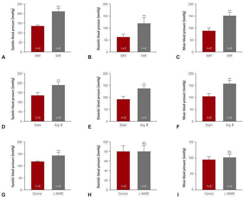
Fig. 1 Elevated blood pressures in hypertensive animal models. (A-C) Systolic, diastolic, and mean blood pressures were measured in a conscious state by the tail-cuff method. Twenty-four-week-old SHR and WKY rats were analyzed (n=8 per group). (D-F) After 2 weeks of angiotensin II (Ang II) infusion in mice, blood pressures were determined (n=9 per group). (G-I) Hypertension was induced by treatment with L-NG-nitro-L-arginine methyl ester (0.1% L-NAME, in drinking water) for 2 weeks, after which, the blood pressures were evaluated. **p<0.01 and ***p<0.001 vs. WKY, sham, or control group. SHR: spontaneously hypertensive rats, WKY: Wistar-Kyoto, NS: not significant, L-NAME: L-NG-nitro-L-arginine methyl ester.
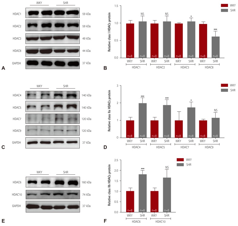
Fig. 2 Class I and IIa/b HDACs expression in spontaneously hypertensive rats. (A) Levels of class I HDACs (HDAC1, 2, 3, and 8) protein expression were determined by western blot analysis using left ventricular tissues from WKY rats and SHR. GAPDH was used as a loading control. (B) Proteins were quantified by densitometry. (C) Levels of cardiac class IIa HDACs (HDAC4, 5, 7, and 9) proteins were assessed in SHR by western blot analysis. (D) Protein levels were quantified by densitometry. (E) Levels of cardiac class IIb HDACs (HDAC6 and HDAC10) proteins were determined by western blot. (F) Quantification of class IIb HDACs. *p<0.05, **p<0.01, and ***p<0.001 vs. WKY. WKY: Wistar-Kyoto, SHR: spontaneously hypertensive rats, HDAC: histone deacetylase, NS: not significant, GAPDH: glyceraldehyde 3-phosphate dehydrogenase.
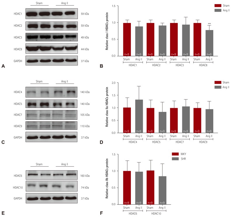
Fig. 3 Decreased expression of HDAC8 in angiotensin II-induced hypertensive mice. (A) Levels of class I HDACs (HDAC1, 2, 3, and 8) protein expression were determined by western blot analysis using hearts from angiotensin II (Ang II)-induced hypertensive mice and sham mice. GAPDH was used as a loading control. (B) Proteins were quantified by densitometry. (C) Levels of cardiac class IIa HDACs (HDAC4, 5, 7, and 9) proteins were assessed in Ang II-induced mice by western blot analysis. (D) Protein levels were quantified by densitometry. (E) Levels of cardiac class IIb HDACs (HDAC6 and HDAC10) proteins were determined by western blot. (F) Quantification of class IIb HDACs. ***p<0.001 vs. sham mice. HDAC: histone deacetylase, GAPDH: glyceraldehyde 3-phosphate dehydrogenase.
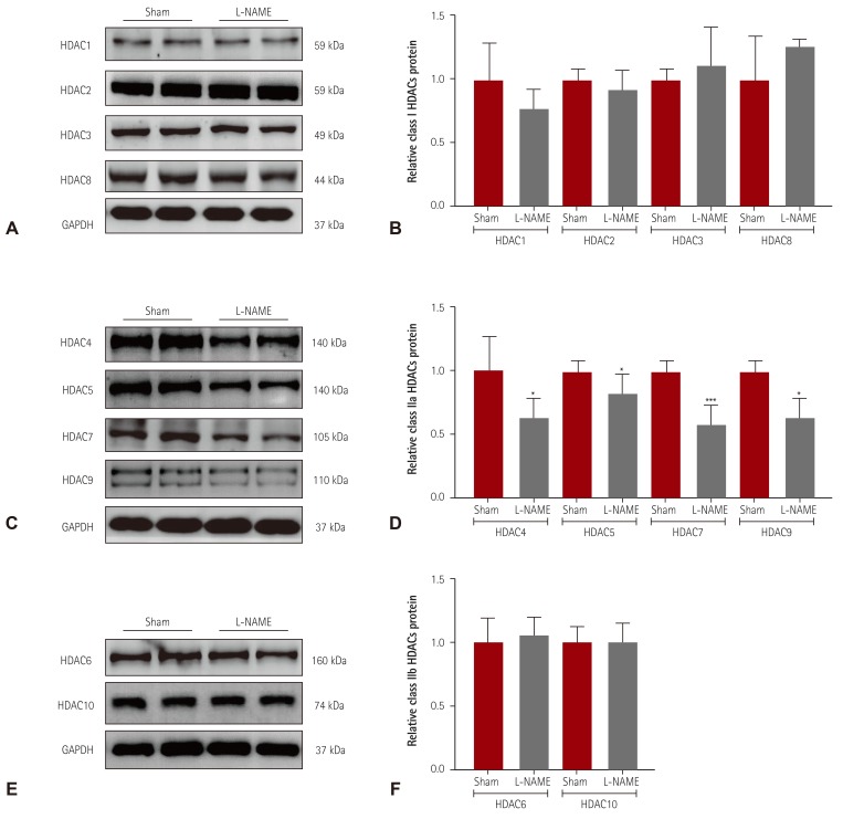
Fig. 4 Decreased class IIa HDAC expression in L-NAME-induced hypertensive mice. (A) Levels of class I HDACs (HDAC1, 2, 3, and 8) protein expression were determined by western blot analysis using hearts from L-NAME-induced hypertensive mice and sham mice. GAPDH was used as a loading control. (B) Proteins were quantified by densitometry. (C) Levels of cardiac class IIa HDACs (HDAC4, 5, 7, and 9) proteins were assessed in L-NAME mice by western blot analysis. (D) Protein levels were quantified by densitometry. (E) Levels of cardiac class IIb HDACs (HDAC6 and HDAC10) were determined by western blot. (F) Quantification of class IIb HDACs. *p<0.05 and ***p<0.001 vs. sham mice. L-NAME: L-NG-nitro-L-arginine methyl ester, GAPDH: glyceraldehyde 3-phosphate dehydrogenase.
Reference
-
1. Ribeiro MO, Antunes E, de Nucci G, Lovisolo SM, Zatz R. Chronic inhibition of nitric oxide synthesis. A new model of arterial hypertension. Hypertension. 1992; 20:298–303. PMID: 1516948.
Article2. Brand S, Amann K, Schupp N. Angiotensin II-induced hypertension dose-dependently leads to oxidative stress and DNA damage in mouse kidneys and hearts. J Hypertens. 2013; 31:333–344. PMID: 23249827.
Article3. Ler SY, Leung CH, Khin LW, et al. HDAC1 and HDAC2 independently predict mortality in hepatocellular carcinoma by a competing risk regression model in a southeast Asian population. Oncol Rep. 2015; 34:2238–2250. PMID: 26352599.
Article4. Koumangoye RB, Andl T, Taubenslag KJ, et al. SOX4 interacts with EZH2 and HDAC3 to suppress microRNA-31 in invasive esophageal cancer cells. Mol Cancer. 2015; 14:24. PMID: 25644061.
Article5. Wilson AJ, Byun DS, Popova N, et al. Histone deacetylase 3 (HDAC3) and other class I HDACs regulate colon cell maturation and p21 expression and are deregulated in human colon cancer. J Biol Chem. 2006; 281:13548–13558. PMID: 16533812.
Article6. Li S, Wang B, Xu Y, Zhang J. Autotaxin is induced by TSA through HDAC3 and HDAC7 inhibition and antagonizes the TSA-induced cell apoptosis. Mol Cancer. 2011; 10:18. PMID: 21314984.
Article7. Lee HA, Lee DY, Cho HM, Kim SY, Iwasaki Y, Kim IK. Histone deacetylase inhibition attenuates transcriptional activity of mineralocorticoid receptor through its acetylation and prevents development of hypertension. Circ Res. 2013; 112:1004–1012. PMID: 23421989.
Article8. Lee HA, Song MJ, Seok YM, Kang SH, Kim SY, Kim I. Histone deacetylase 3 and 4 complex stimulates the transcriptional activity of the mineralocorticoid receptor. PLoS One. 2015; 10:e0136801. PMID: 26305553.
Article9. Usui T, Okada M, Mizuno W, et al. HDAC4 mediates development of hypertension via vascular inflammation in spontaneous hypertensive rats. Am J Physiol Heart Circ Physiol. 2012; 302:H1894–H1904. PMID: 22389387.
Article10. Kameshima S, Okada M, Yamawaki H. Expression and localization of calmodulin-related proteins in brain, heart and kidney from spontaneously hypertensive rats. Biochem Biophys Res Commun. 2016; 469:654–658. PMID: 26697749.
Article11. Cavasin MA, Demos-Davies K, Horn TR, et al. Selective class I histone deacetylase inhibition suppresses hypoxia-induced cardiopulmonary remodeling through an antiproliferative mechanism. Circ Res. 2012; 110:739–748. PMID: 22282194.
Article12. Pang J, Yan C, Natarajan K, et al. GIT1 mediates HDAC5 activation by angiotensin II in vascular smooth muscle cells. Arterioscler Thromb Vasc Biol. 2008; 28:892–898. PMID: 18292392.
Article13. Kee HJ, Bae EH, Park S, et al. HDAC inhibition suppresses cardiac hypertrophy and fibrosis in DOCA-salt hypertensive rats via regulation of HDAC6/HDAC8 enzyme activity. Kidney Blood Press Res. 2013; 37:229–239. PMID: 23868068.
Article14. Lemon DD, Horn TR, Cavasin MA, et al. Cardiac HDAC6 catalytic activity is induced in response to chronic hypertension. J Mol Cell Cardiol. 2011; 51:41–50. PMID: 21539845.
Article15. Choi SY, Ryu Y, Kee HJ, et al. Tubastatin A suppresses renal fibrosis via regulation of epigenetic histone modification and Smad3-dependent fibrotic genes. Vascul Pharmacol. 2015; 72:130–140. PMID: 25921924.
Article16. Kee HJ, Kwon JS, Shin S, Ahn Y, Jeong MH, Kook H. Trichostatin A prevents neointimal hyperplasia via activation of Krüppel like factor 4. Vascul Pharmacol. 2011; 55:127–134. PMID: 21763782.
Article17. Xu X, Ha CH, Wong C, et al. Angiotensin II stimulates protein kinase D-dependent histone deacetylase 5 phosphorylation and nuclear export leading to vascular smooth muscle cell hypertrophy. Arterioscler Thromb Vasc Biol. 2007; 27:2355–2362. PMID: 17823368.
Article18. Usui T, Morita T, Okada M, Yamawaki H. Histone deacetylase 4 controls neointimal hyperplasia via stimulating proliferation and migration of vascular smooth muscle cells. Hypertension. 2014; 63:397–403. PMID: 24166750.
Article19. Kim GR, Cho SN, Kim HS, et al. HDAC4 and GATA6 regulate arterial remodeling in angiotensin ii-induced hypertension. J Hypertens. 2016; 34:2206–2219. PMID: 27512969.20. Baylis C, Mitruka B, Deng A. Chronic blockade of nitric oxide synthesis in the rat produces systemic hypertension and glomerular damage. J Clin Invest. 1992; 90:278–281. PMID: 1634615.
Article21. Bartunek J, Weinberg EO, Tajima M, et al. Chronic N(G)-nitro-L-arginine methyl ester-induced hypertension: novel molecular adaptation to systolic load in absence of hypertrophy. Circulation. 2000; 101:423–429. PMID: 10653835.
- Full Text Links
-
- Actions
-
Cited
- CITED
-
- Close
- Share
-
- Similar articles
-
- Leptomycin B Increases Radiosensitization by Trichostain A in HeLa Cells
- Transcription of class II MHC gene by interferon-gamma in FRTL-5 cells
- A study of the etiology of unilateral Class II, division 1 malocclusion
- Comparison of occlusal contact areas of class I and class II molar relationships at finishing using three-dimensional digital models
- Epidemiologic study of the prevalence of malocclusion in Korean
