Clin Endosc.
2017 Mar;50(2):209-210. 10.5946/ce.2016.041.
Nodular Lymphoid Hyperplasia with Aggressive Endoscopic Appearance in the Colon of an Adult Woman
- Affiliations
-
- 1Department of Gastroenterology, Benizelion General Hospital, Heraklion, Greece. gpaspatis@gmail.com
- 2Department of Pathology, Benizelion General Hospital, Heraklion, Greece.
- 3Department of Surgery, Benizelion General Hospital, Heraklion, Greece.
- KMID: 2383541
- DOI: http://doi.org/10.5946/ce.2016.041
Abstract
- No abstract available.
MeSH Terms
Figure
-
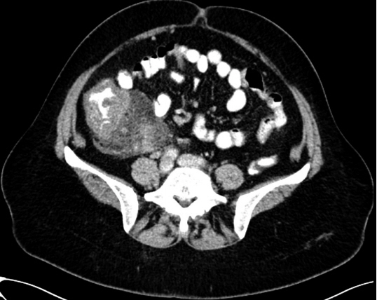
Fig. 1. Computed tomography scan showing circular wall thickening in the ascending colon, and the terminal ileum with enlarged lymph nodes.
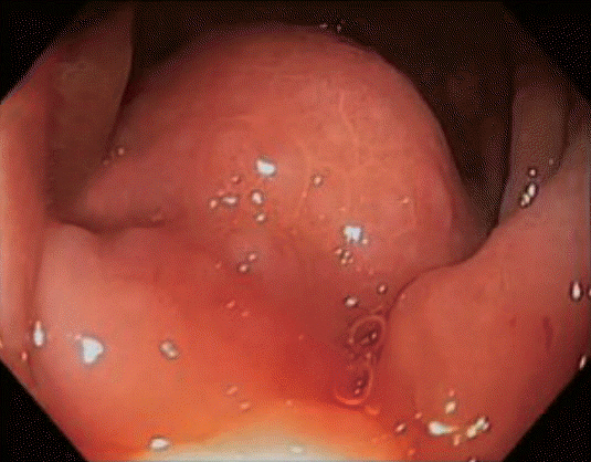
Fig. 2. Protruding mass in the hepatic flexure.

Fig. 3. Section from the cecum wall (H&E stain, ×20), showing prominent lymphoid follicles, with highly reactive germinal centers, located in the mucosa and submucosa, suggesting nodular lymphoid hyperplasia.
Reference
-
1. Albuquerque A. Nodular lymphoid hyperplasia in the gastrointestinal tract in adult patients: a review. World J Gastrointest Endosc. 2014; 6:534–540.
Article2. Ranchod M, Lewin KJ, Dorfman RF. Lymphoid hyperplasia of the gastrointestinal tract. A study of 26 cases and review of the literature. Am J Surg Pathol. 1978; 2:383–400.3. Rubio-Tapia A, Hernández-Calleros J, Trinidad-Hernández S, Uscanga L. Clinical characteristics of a group of adults with nodular lymphoid hyperplasia: a single center experience. World J Gastroenterol. 2006; 12:1945–1948.
Article4. Chandra S. Benign nodular lymphoid hyperplasia of colon: a report of two cases. Indian J Gastroenterol. 2003; 22:145–146.
- Full Text Links
-
- Actions
-
Cited
- CITED
-
- Close
- Share
-
- Similar articles
-
- A Case of Primary Jejunal Malignant Lymphoma Associated with Nodular Lymphoid Hyperplasia
- Surgical Treatment of the Pulmonary Nodular Lymphoid Hyperplasia: A case report
- Diagnostic Pediatric Colonoscopy for Lymphoid Hyperplasia of Terminal Ileum
- Endoscopic features and clinicopathological relationship of colonic lymphoid hyperplasia
- Pulmonary Nodular Lymphoid Hyperplasia in a 33-Year-Old Woman
