Non-umbilical Cutaneous Metastasis of Pancreatic Adenocarcinoma as the First Clinical Manifestation: A Case Report
- Affiliations
-
- 1Department of Internal Medicine, Sanggye Paik Hospital, Inje University College of Medicine, Seoul, Korea. human@paik.ac.kr
- 2Department of Pathology, Sanggye Paik Hospital, Inje University College of Medicine, Seoul, Korea.
- KMID: 2383520
- DOI: http://doi.org/10.4166/kjg.2016.68.4.221
Abstract
- Non-umbilical cutaneous metastases from pancreatic adenocarcinomas are extremely rare. Only a few cases have been reported in the literature. An 83-year-old Korean woman, with no previous medical history, presented with a painful nodule on her scalp. Histologic examination of the nodule revealed a metastatic adenocarcinoma, and immunohistochemical staining was positive for cytokeratin (CK) 7 and CK 19. These findings were consistent with a metastatic carcinoma of pancreatic origin. An abdominal computed tomography scan identified a mass on the pancreatic head and multiple enlarged lymph nodes. Pathological examination of an endoscopic ultrasound-guided fine needle biopsy of the pancreatic mass determined that it was a poorly differentiated carcinoma. The patient refused any treatment owing to her old age and short life expectancy. Four months later, the disease progressed rapidly, and the patient died.
MeSH Terms
Figure
-
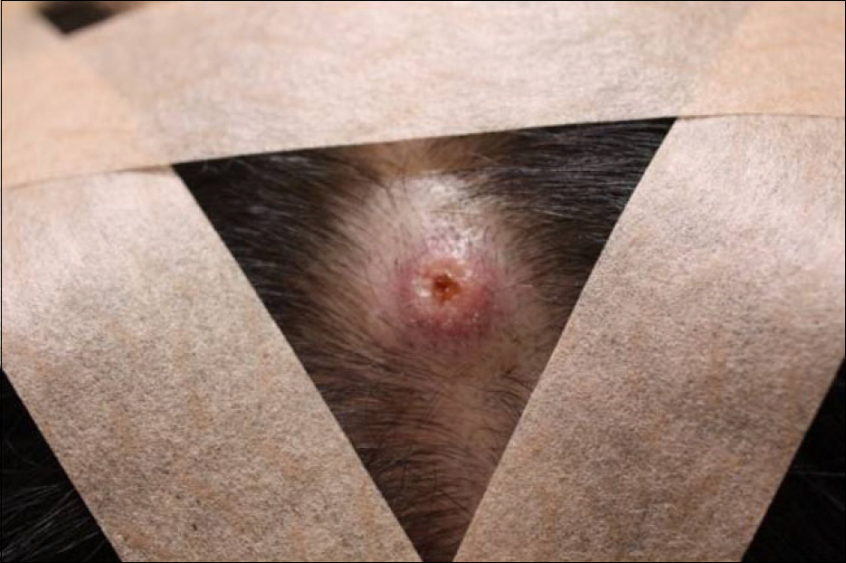
Fig. 1. Erythematous plaque with a central plug is seen on the scalp.
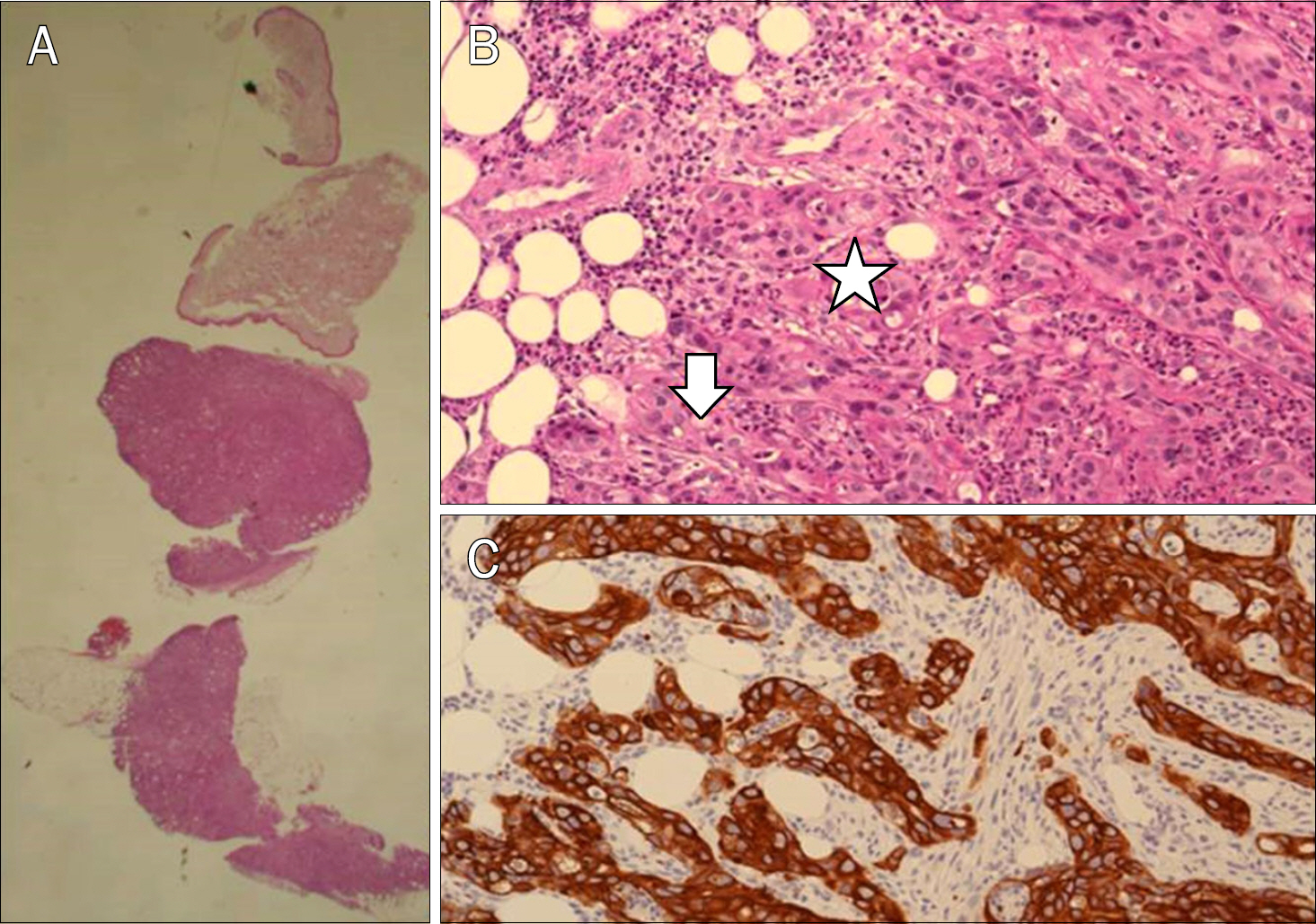
Fig. 2. (A) Histology section of the scalp lesion. The nodule is highly cellular and infiltrating towards the fat lobule. (B) Ductal proliferation with severe nuclear atypia, frequent mitoses, intraluminal necrotic debris (asterisk), and intracytoplasmic mucin vacuoles (arrow), suggestive of high-grade adenocarcinoma. (C) Diffuse and strong staining for cytokeratin 7 in the tumor cells, supportive of a ductal type carcinoma (such as pancreas, breast, or biliary).
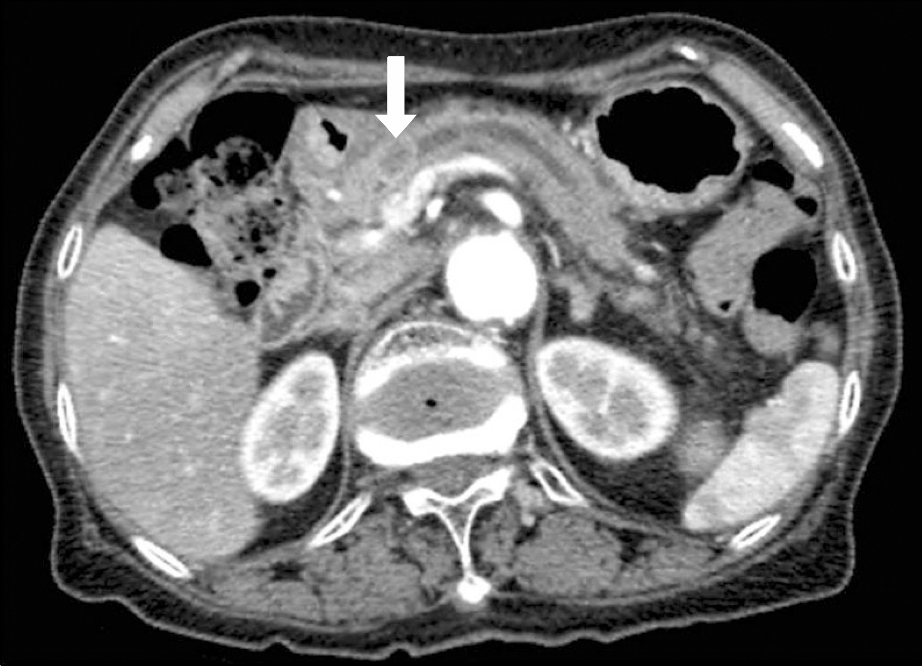
Fig. 3. Abdominal CT scan shows a 1.4-cm, ill-defined, ovoid, low-density lesion (arrow) in the pancreatic head with upstream pancreatic duct dilatation.
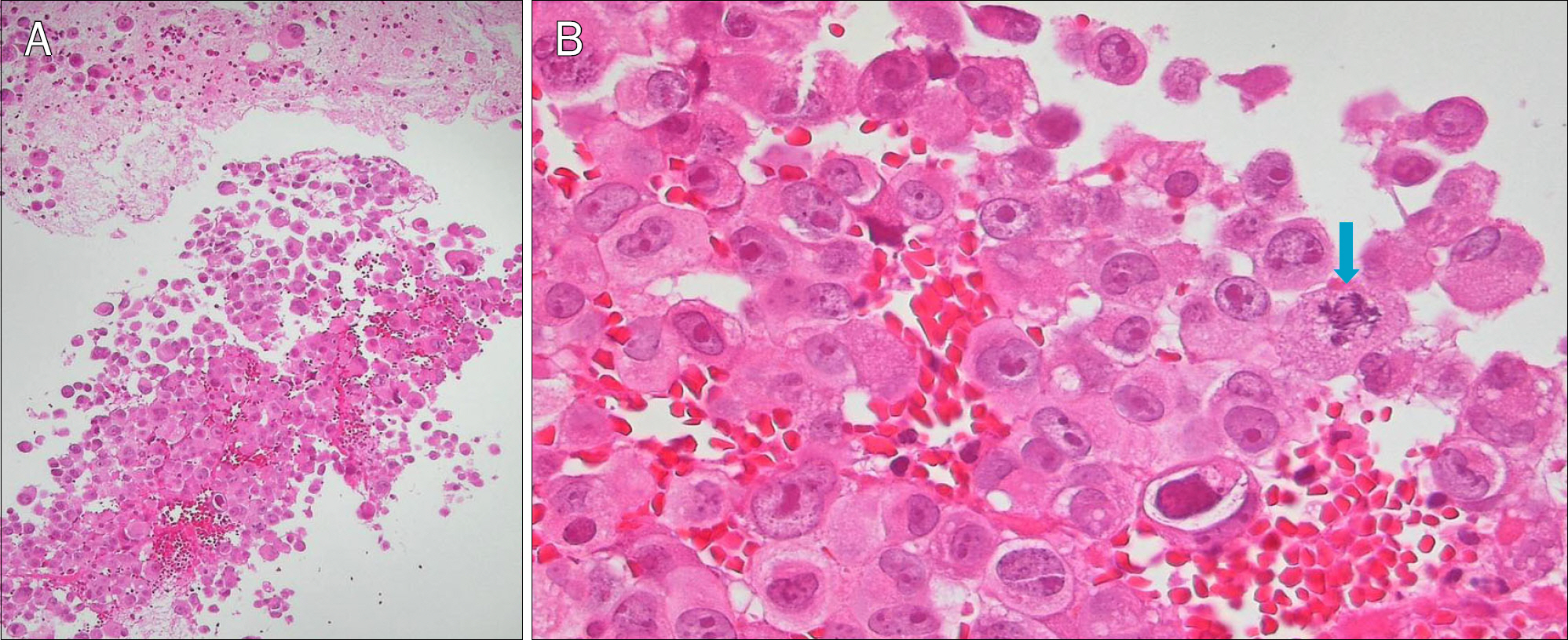
Fig. 4. (A) The needle biopsy from the pancreatic head mass shows loosely cohesive atypical epithelial cells, embedded in a blood-tinged fibrin clot (H&E, ×40). (B) Pleomorphic tumor cells with vesicular nuclei and several prominent nucleoli. Occasional atypical mitoses are indicated (arrow) (H&E, ×400).
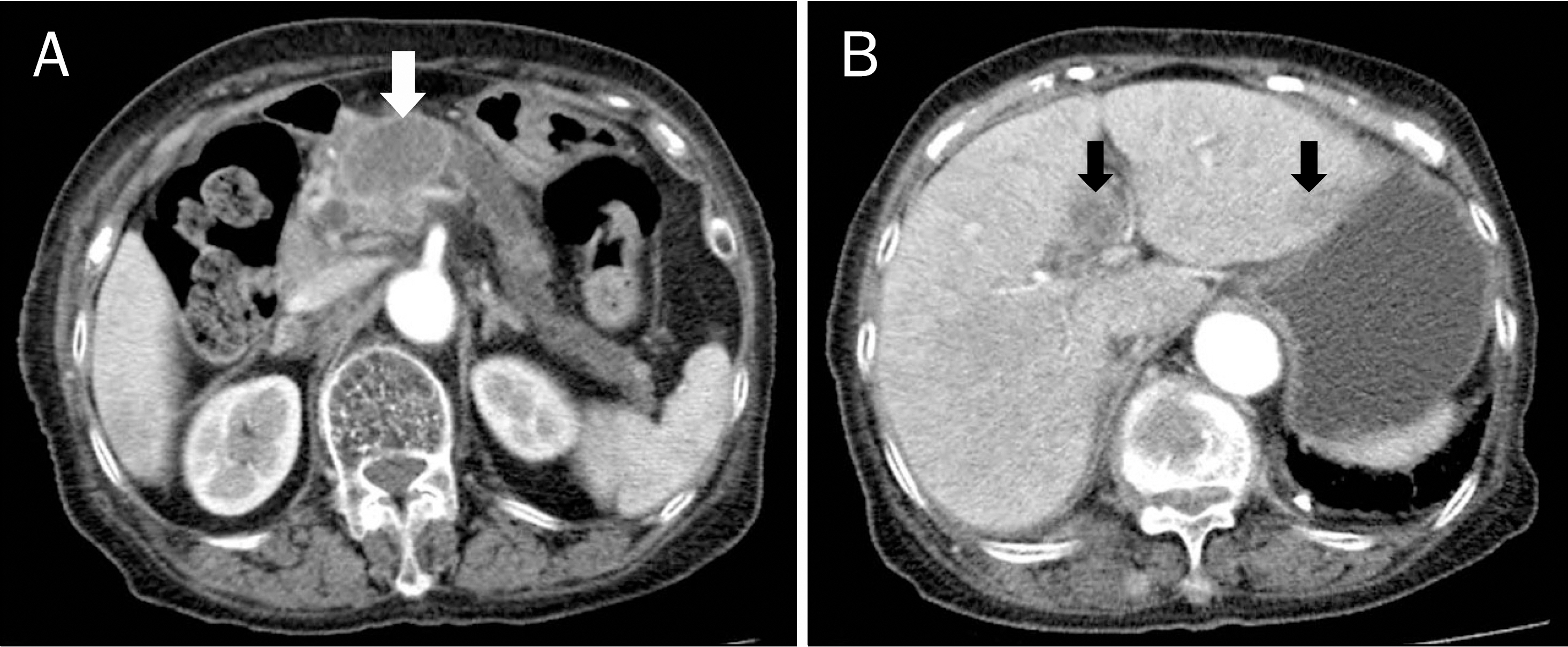
Fig. 5. (A) Abdominal CT scan taken three months after the diagnosis shows the increased size (4.1 cm) of the pancreatic head mass with upstream pancreatic duct dilatation (white arrow). (B) Multiple newly detected metastases to the liver (black arrows).
Cited by 1 articles
-
Clinicopathologic features of cutaneous metastases from internal malignancies
Hyeong Mok Kwon, Gyu Yeong Kim, Dong Hoon Shin, Young Kyung Bae
J Pathol Transl Med. 2021;55(4):289-297. doi: 10.4132/jptm.2021.05.24.
Reference
-
References
1. Zhou HY, Wang XB, Gao F, Bu B, Zhang S, Wang Z. Cutaneous metastasis from pancreatic cancer: a case report and systematic review of the literature. Oncol Lett. 2014; 8:2654–2660.
Article2. Kaoutzanis C, Chang MC, Abdul Khalek FJ, Kreske E. Non-um-bilical cutaneous metastasis of a pancreatic adenocarcinoma. BMJ Case Rep. 2013; 2013:bcr2012007931.
Article3. Bdeiri K, Kamar FG. Cutaneous metastasis of pancreatic adenocarcinoma as a first clinical manifestation: a case report and review of the literature. Gastrointest Cancer Res. 2013; 6:61–63.4. van Akkooi AC, Dokter J, Boxma H. Unusual first presentation of metastatic pancreatic cancer as skin metastases in a burn patient. Burns. 2010; 36:e111–e114.
Article5. Jun DW, Lee OY, Park CK, et al. Cutaneous metastases of pancreatic carcinoma as a first clinical manifestation. Korean J Intern Med. 2005; 20:260–263.
Article6. Lim D, Ha M, Song I. Trends in major cancer mortality in Korea, 1983–2012, with a joinpoint analysis. Cancer Epidemiol. 2015; 39:939–946.
Article7. Pontinen T, Melin A, Varadi G, et al. Cutaneous metastasis of pancreatic adenocarcinoma after kidney transplant: a case report and review of the literature. Exp Clin Transplant. 2010; 8:273–276.8. Yendluri V, Centeno B, Springett GM. Pancreatic cancer presenting as a Sister Mary Joseph's nodule: case report and update of the literature. Pancreas. 2007; 34:161–164.9. Abdel-Hafez HZ. Cutaneous pancreatic metastasis: a case report and review of literature. Dermatol Surg. 2008; 34:1580–1583.
Article10. Gawrieh S, Massey BT, Komorowski RA. Scalp metastases as the first manifestation of pancreatic cancer. Dig Dis Sci. 2002; 47:1469–1471.11. Nakano S, Narita R, Yamamoto M, Ogami Y, Osuki M. Two cases of pancreatic cancer associated with skin metastases. Am J Gastroenterol. 1996; 91:410–411.12. Taniguchi S, Hisa T, Hamada T. Cutaneous metastases of pancreatic carcinoma with unusual clinical features. J Am Acad Dermatol. 1994; 31:877–880.
Article13. Galvañ VG. Sister Mary Joseph's nodule. Ann Intern Med. 1998; 128:410.
Article14. Takemura N, Fujii N, Tanaka T. Cutaneous metastasis as the first clinical manifestation of pancreatic adenocarcinoma: a case treated with gemcitabine. J Dermatol. 2007; 34:662–664.
Article15. Takeuchi H, Kawano T, Toda T, et al. Cutaneous metastasis from pancreatic adenocarcinoma: a case report and a review of the literature. Hepatogastroenterology. 2003; 50:275–277.16. Brownstein MH, Helwig EB. Patterns of cutaneous metastasis. Arch Dermatol. 1972; 105:862–868.
Article17. Matros E, Bailey G, Clancy T, et al. Cytokeratin 20 expression identifies a subtype of pancreatic adenocarcinoma with decreased overall survival. Cancer. 2006; 106:693–702.
Article18. Lookingbill DP, Spangler N, Sexton FM. Skin involvement as the presenting sign of internal carcinoma. A retrospective study of 7316 cancer patients. J Am Acad Dermatol. 1990; 22:19–26.
- Full Text Links
-
- Actions
-
Cited
- CITED
-
- Close
- Share
-
- Similar articles
-
- A Case of Cutaneous Metastatic Adenosquamous Carcinoma of the Pancreas
- Non-umbilical Cutaneous Metastasis of Pancreatic Adenocarcinoma
- A Case of Cutaneous Metastasis from Pancreatic Adenocarcinoma
- A Case of Umbilical Metastasis as the Presenting Sign of Pancreatic Adenocarcinoma
- Cutaneous Metastases of Pancreatic Carcinoma as a First Clinical Manifestation
