Duodenal Loop Obstruction as an Unusual Cause of Acute Pancreatitis: A Case Series
- Affiliations
-
- 1Department of Internal Medicine, Chungbuk National University College of Medicine, Cheongju, Korea. smpark@chungbuk.ac.kr
- KMID: 2383464
- DOI: http://doi.org/10.4166/kjg.2016.68.6.326
Abstract
- Duodenal loop obstruction is an unusual cause of acute pancreatitis. Increased intraluminal pressure hinders pancreatic flow, causing dilatation of the pancreatic duct and inducing acute pancreatitis. We experienced three cases of acute pancreatitis that resulted from duodenal loop obstruction after (1) an esophagectomy with gastric pull-up procedure for esophageal cancer, (2) a gastrectomy with Billroth I reconstruction for gastric cancer, and (3) a gastrojejunostomy for abdominal trauma. An abdominal CT scan revealed a distended duodenal loop, dilated pancreatic duct, and inflamed pancreas with fluid collection. Acute pancreatitis with duodenal loop obstruction was diagnosed by abdominal pain, elevated serum amylase/lipase, and abdominal CT findings. Immediate decompression with a nasogastric tube was performed, and all patients showed improvement within one week after admission. Each patient was followed up for more than two years without recurrence. Our findings suggest the usefulness of nasogastric tube decompression as the first line of treatment for acute pancreatitis related to duodenal loop obstruction.
MeSH Terms
Figure
-
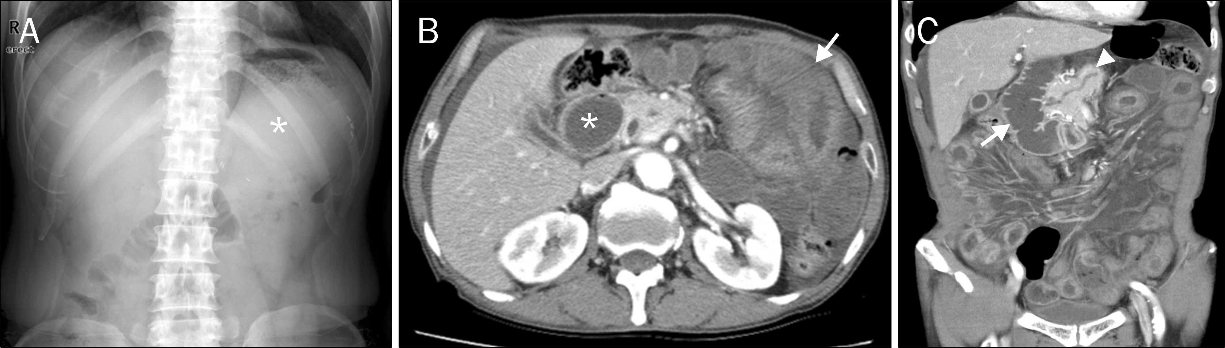
Fig. 1. Plain abdomen and abdominal CT images of Case 1. (A) Suspicious distended stomach filled with fluid (asterisk) without abnormal bowel gas. (B) Axial view. Dilatation of the duodenum (asterisk) and diffuse small bowel wall thickening (arrow). (C) Coronal view. Distended duodenal loop (arrow) and mild pancreatic duct dilation (arrowhead).
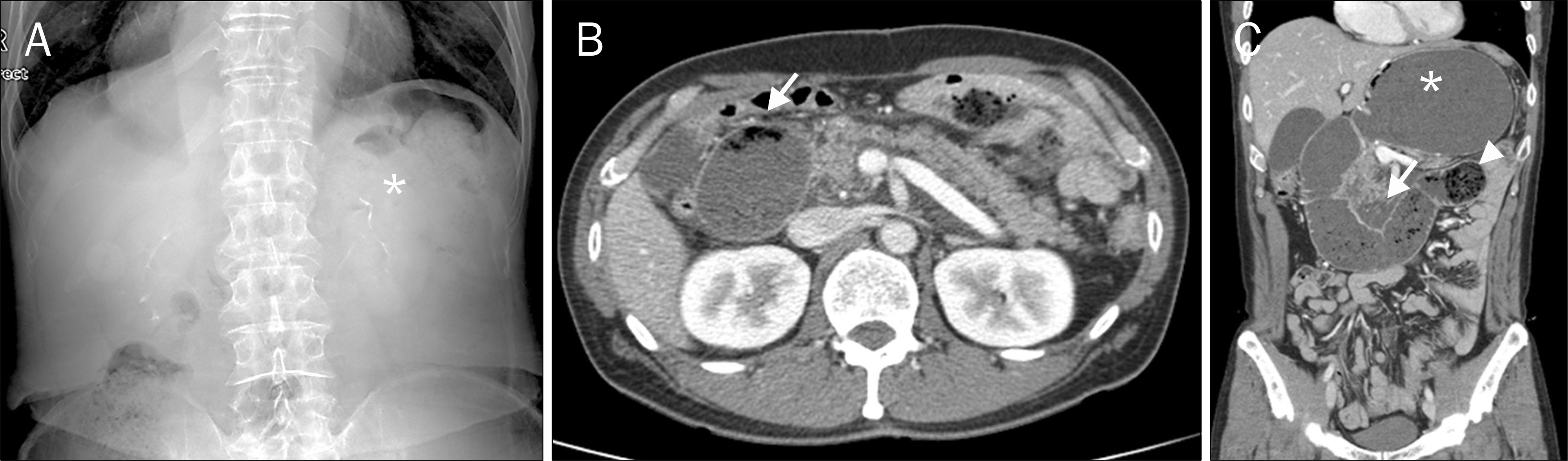
Fig. 2. Plain abdomen and abdominal CT images of Case 2. (A) Suspicious distended stomach filled with fluid (asterisk) without abnormal bowel gas. (B) Axial view. A massive duodenal dilatation with a large volume of food inside (arrow). (C) Coronal view. Dilatation of the stomach (asterisk) and duodenal loop with abrupt luminal narrowing of the proximal jejunum (arrowhead). Swelling of the pancreas with peripancreatic fluid collection (arrow) is suggestive of acute pancreatitis.
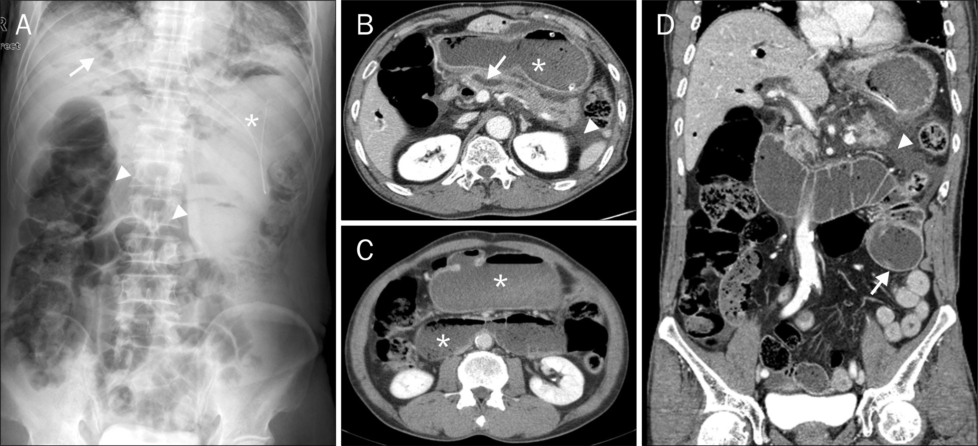
Fig. 3. Plain abdomen and abdominal CT images of Case 3. (A) Linear air at right upper quadrant area (arrow), suspicious fluid-filled stomach (asterisks), and gaseous distended colonic loop and small bowels at right sided abdomen (arrowheads). (B, C) Axial views. Marked dilation of the stomach and duodenum (asterisks) and dilation of the pancreatic duct (arrow) with fluid collection (arrowhead). (D) Coronal view. A distended duodenal loop with abrupt luminal narrowing of the proximal jejunum (arrow) and fluid collection at the pancreatic tail (arrowhead). Plain abdominal film was revealed.
Reference
-
References
1. Vettoretto N, Pettinato G, Romessis M, Bravo AF, Barozzi G, Giovanetti M. Laparoscopy in afferent loop obstruction presenting as acute pancreatitis. JSLS. 2006; 10:270–274.2. Lankisch PG, Petersen F, Brinkmann G. An enormous ventral (epigastric) hernia as a cause of acute pancreatitis: Pfeffer's closed duodenal loop model in the animal, first seen in a human. Gastroenterology. 2003; 124:865–866.
Article3. Korrapati V, Sidhu-Buonocore S, Grendell JH. Acute pancreatitis and pseudocyst due to a closed loop obstruction from an epigastric hernia. Clin Gastroenterol Hepatol. 2009; 7:e48.
Article4. Joo YE, Kim HS, Choi SK, et al. Internal hernia presenting as obstructive jaundice and acute pancreatitis. Scand J Gastroenterol. 2002; 37:983–986.
Article5. Kim HH, Park SJ, Park MI, Moon W. Acute gastric dilatation and acute pancreatitis in a patient with an eating disorder: solving a chicken and egg situation. Intern Med. 2011; 50:571–575.
Article6. Frick TW, Schretzenmaier M, Hoffmann R, Largiadèr F. Obstructive ileus and acute pancreatitis. Z Gastroenterol. 1990; 28:206–207.7. De Martino C, Caiazzo P, Albano M, et al. Acute afferent loop obstruction treated by endoscopic decompression. Case report and review of literature. Ann Ital Chir. 2012; 83:555–558.8. Kim HJ, Kim JW, Kim KH, et al. A case of afferent loop syndrome treated by endoscopic drainage procedure using nasogastric tube. Korean J Gastroenterol. 2007; 49:173–176.9. Kitamura H, Miwa S, Nakata T, et al. Sonographic detection of visceral adhesion in percutaneous drainage of afferent-loop small-intestine obstruction. J Clin Ultrasound. 2000; 28:133–136.
Article10. Burdick JS, Garza AA, Magee DJ, Dykes C, Jeyarajah R. Endoscopic management of afferent loop syndrome of malignant etiology. Gastrointest Endosc. 2002; 55:602–605.
Article11. Alawneh I. Afferent loop obstruction after gastrectomy simulating acute pancreatitis. Int Surg. 1980; 65:415–417.12. Inoue M, Uchida K, Otake K, Okigami M, Maji T, Kusunoki M. Development of acute pancreatitis after Nissen fundoplication. Pediatr Int. 2015; 57:e48–e49.
Article13. Pfeffer RB, Stasior O, Hinton JW. The clinical picture of the sequential development of acute hemorrhagic pancreatitis in the dog. Surg Forum. 1957; 8:248–251.14. Ferrie MM, O'Hare R, Joffe SN. Acute and chronic pancreatitis in the rat caused by a closed duodenal loop. Digestion. 1978; 18:280–285.
Article15. Sugimoto M, Takada T, Yasuda H. A new experimental pancreatitis by incomplete closed duodenal loop: the influence of pancreatic microcirculation on the development and progression of induced severe pancreatitis in rats. Pancreas. 2004; 28:e112–e129.16. Davis S, Parbhoo SP, Gibson MJ. The plain abdominal radiograph in acute pancreatitis. Clin Radiol. 1980; 31:87–93.
Article
- Full Text Links
-
- Actions
-
Cited
- CITED
-
- Close
- Share
-
- Similar articles
-
- Acute Abdomen Caused by Complicated Afferent Loop Syndrome after Gastrectomy
- Successful Endoscopic Decompression for Intramural Duodenal Hematoma with Gastric Outlet Obstruction Complicating Acute Pancreatitis
- Recurrent Pancreatitis Caused by Afferent Loop Syndrome with Pathologic Features of Type II Autoimmune Pancreatitis
- A Case of Acute Gastric Dilatation with Duodenal Obstruction Accompanying Elevated Serum Pancreatic Enzymes in a Patient with Bulimia
- A Case of Pancreatitis-Induced Intramural Duodenal Hematoma
