Bowenoid Papulosis of the Vulva and Subsequent Periungual Bowen's Disease Induced by the Same Mucosal HPVs
- Affiliations
-
- 1Department of Dermatology, School of Medicine, Pusan National University, Busan, Korea. drkmp@hanmail.net
Abstract
- We report the case of a 23-year-old woman who developed bowenoid papulosis of the vulva and subsequent periungual Bowen's disease. She had a history of a long standing periungual wart on her right thumb before the outbreak of periungual Bowen's disease. By HPV DNA chip, human papillomavirus (HPV) 11, 18 and 31 were identified from the periungual lesions, and HPV 11, 18 and 33 from the vulvar lesion. This case supports the theory of anogenital-digital spread of HPV, and proposes that the periungual wart may change into Bowen's disease by mucosal HPVs. To the best of our knowledge, this case is important as the first Korean case of periungual Bowen's disease concurrent with bowenoid papulosis of the vulva.
Keyword
MeSH Terms
Figure
-

Fig. 1 (A) Multiple pigmented verrucous papules and plaques on the periungual area for 4 months. (B) Hyperpigmented verrucous plaques on the genital area for 7 months.
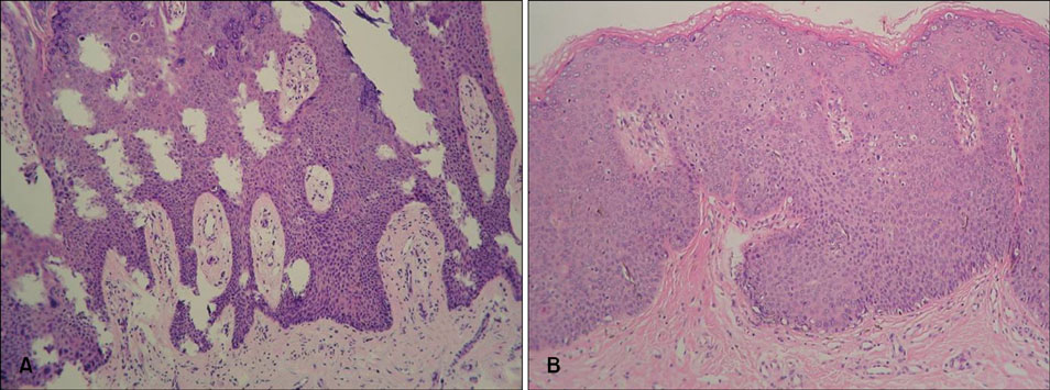
Fig. 2 (A) The biopsy specimen from the periungual lesion shows severe cytologic atypia (nuclear enlargement, hyperchromasia, and pleomorphism), numerous mitoses, and scattered dyskeratotic cells (H&E, ×100). (B) The biopsy specimen from the vulvar lesion shows full-thickness atypia and dyskeratosis (H&E, ×100).
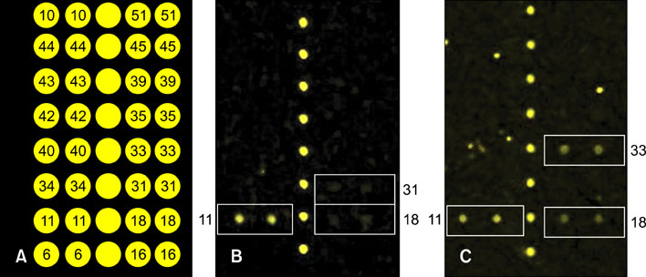
Fig. 3 The HPV DNA chip was amplified from the human papillomavirus (HPV) polymerase chain reaction and subsequently hybridized to oligonucleotide probes specific to each genotype. (A) The positive signal was represented as double spots. (B) Combined infections of HPV type 11, 18 and 31 in periungual lesion. (C) Combined infections of HPV type 11, 18 and 33 in vulvar lesion.
Reference
-
1. Kreuter A, Gambichler T, Pfister H, Wieland U. Diversity of human papillomavirus types in periungual squamous cell carcinoma. Br J Dermatol. 2009. 161:1262–1269.
Article2. Meyer T, Arndt R, Christophers E, Nindl I, Stockfleth E. Importance of human papillomaviruses for the development of skin cancer. Cancer Detect Prev. 2001. 25:533–547.3. Alam M, Caldwell JB, Eliezri YD. Human papillomavirus-associated digital squamous cell carcinoma: literature review and report of 21 new cases. J Am Acad Dermatol. 2003. 48:385–393.
Article4. Ibe M, Kawase M, Ishiji T, Kamide R, Niimura M. A cardiac allograft recipient with Bowen's disease on a finger and concurrent perianal bowenoid papulosis. J Dermatol. 2003. 30:389–394.
Article5. Nordin P, Stenquist B, Hansson BG. Joint occurrence of human papillomavirus type 16 DNA in Bowen's disease on a finger and in dysplasia of the vulva and the uterine cervix. Br J Dermatol. 1994. 131:740.
Article6. Hara H, Honda A, Suzuki H, Sata T, Matsukura T. Detection of human papillomavirus type 58 in polydactylous Bowen's disease on the fingers and toes of a woman - concurrent occurrence of invasive vulval and cervical carcinomas. Dermatology. 2004. 209:218–222.
Article7. Forslund O, Nordin P, Hansson BG. Mucosal human papillomavirus types in squamous cell carcinomas of the uterine cervix and subsequently on fingers. Br J Dermatol. 2000. 142:1148–1153.
Article
- Full Text Links
-
- Actions
-
Cited
- CITED
-
- Close
- Share
-
- Similar articles
-
- A Study on the Histopathologic Features of Bowenoid Papulosis and the Numerical Change in Langerhans Cells
- A Case of Bowenoid Papulosis
- Invasive carcinoma of the vulva arising from bowenoid papulosis: A case report
- Pagetoid Bowen's Disease on the Vulva
- A Case of Bowenoid Papulosis Occurred in Genital Warts
