J Korean Neurosurg Soc.
2017 May;60(3):371-374. 10.3340/jkns.2015.0708.010.
Giant Ganglioneuroma of Thoracic Spine: A Case Report and Review of Literature
- Affiliations
-
- 1Department of Orthopedics, The First Hospital of Jilin University, Changchun, China. haungyong18@163.com
- 2Department of Orthopedics, Taihe Hospital, Hubei University of Medicine, Shiyan, China.
- KMID: 2382774
- DOI: http://doi.org/10.3340/jkns.2015.0708.010
Abstract
- Ganglioneuroma (GN) is a rare benign tumor of neural crest origin usually found in the abdomen, but may occasionally present at uncommon sites including the cervical, lumbar, or sacral spine. However, GNs of thoracic spine are extremely rare. In this report, we describe a 12-year-old girl with giant GN in the thoracic spine, who underwent successful resection (T1-4 level) of the tumor. Histopathological examination confirmed the diagnosis. GN should be considered in the differential diagnosis of any paraspinal mass. A high index of suspicion and correlation of clinico-radiological findings is necessary in differentiating a large benign tumor from a malignant growth. Complete surgical excision is the treatment of choice; however tumor size and location need to be considered for the surgical approach (one-step or multiple surgeries). Close follow-up after surgery is mandatory.
Keyword
MeSH Terms
Figure
-
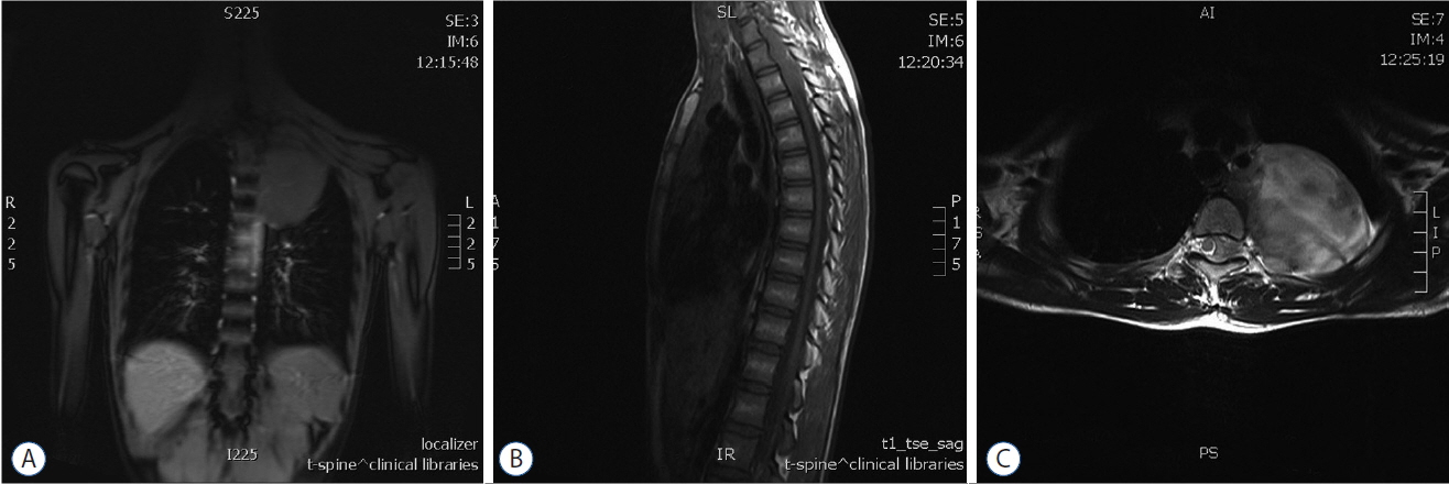
Fig. 1 Preoperative MRI. A: Coronal T1-weighted image showing a giant, mildly hypointense left-sided mass. B: Sagittal T1-weighted MRI showing a hypointense strip at the T1–T4 level. C: Axial T2-weighted MRI at the T3–4 level showing a heterogeneously hyper-intense mass extending to the spinal canal. MRI: magnetic resonance imaging.

Fig. 2 H&E stained section showing mature ganglion cells surrounded by schwann cells and fibrillary stroma (400×).
Reference
-
References
1. Hioki A, Miyamoto K, Hirose Y, Kito Y, Fushimi K, Shimizu K. Cervical symmetric dumbbell ganglioneuromas causing severe paresis. Asian Spine J. 8:74–78. 2014.
Article2. Kara T, Oztunali C. Radiologic findings of thoracic scoliosis due to giant ganglioneuroma. Clin Imaging. 37:767–768. 2013.
Article3. Kyoshima K, Sakai K, Kanaji M, Oikawa S, Kobayashi S, Sato A, et al. Symmetric dumbbell ganglioneuromas of bilateral C2 and C3 roots with intradural extension associated with von Recklinghausen’s disease: case report. Surg Neurol. 61:468–473. discussion 473. 2004.
Article4. Pang BC, Tchoyoson Lim CC, Tan KK. Giant spinal ganglioneuroma. J Clin Neurosci. 12:967–972. 2005.
Article5. Son DW, Song GS, Kim YH, Lee SW. Ventrally located cervical dumbbell ganglioneuroma producing spinal cord compression. Korean J Spine. 10:246–248. 2013.
Article6. Tsai FJ, Kuo KL, Tzou RD, Cheng YH, Hwang SL, Lieu AS. A huge extradural ganglioneuroma of the lumbar spine. Formosan J Surg. 47:160–165. 2014.
Article7. Ugarriza LF, Cabezudo JM, Ramirez JM, Lorenzana LM, Porras LF. Bilateral and symmetric C1–C2 dumbbell ganglioneuromas producing severe spinal cord compression. Surg Neurol. 55:228–231. 2001.
Article8. Vardas K, Manganas D, Papadimitriou G, Vougas V, Bakalis A, Chantziara M, et al. Presacral ganglioneuroma: diagnostic considerations and therapeutic strategy. Case Rep Oncol. 6:561–568. 2013.
Article
