A Case of Primary Subpleural Pulmonary Microcystic Myxoma Coincidentally Occurred with Pulmonary Adenocarcinoma
- Affiliations
-
- 1Department of Pathology, Gachon University Gil Medical Center, Incheon, Korea. clara_nrk@gilhospital.com
- 2Department of Thoracic Surgery, Gachon University Gil Medical Center, Incheon, Korea.
- 3Department of Radiology, Gachon University Gil Medical Center, Incheon, Korea.
- KMID: 2381389
- DOI: http://doi.org/10.4132/jptm.2015.03.12
Abstract
- No abstract available.
MeSH Terms
Figure
-
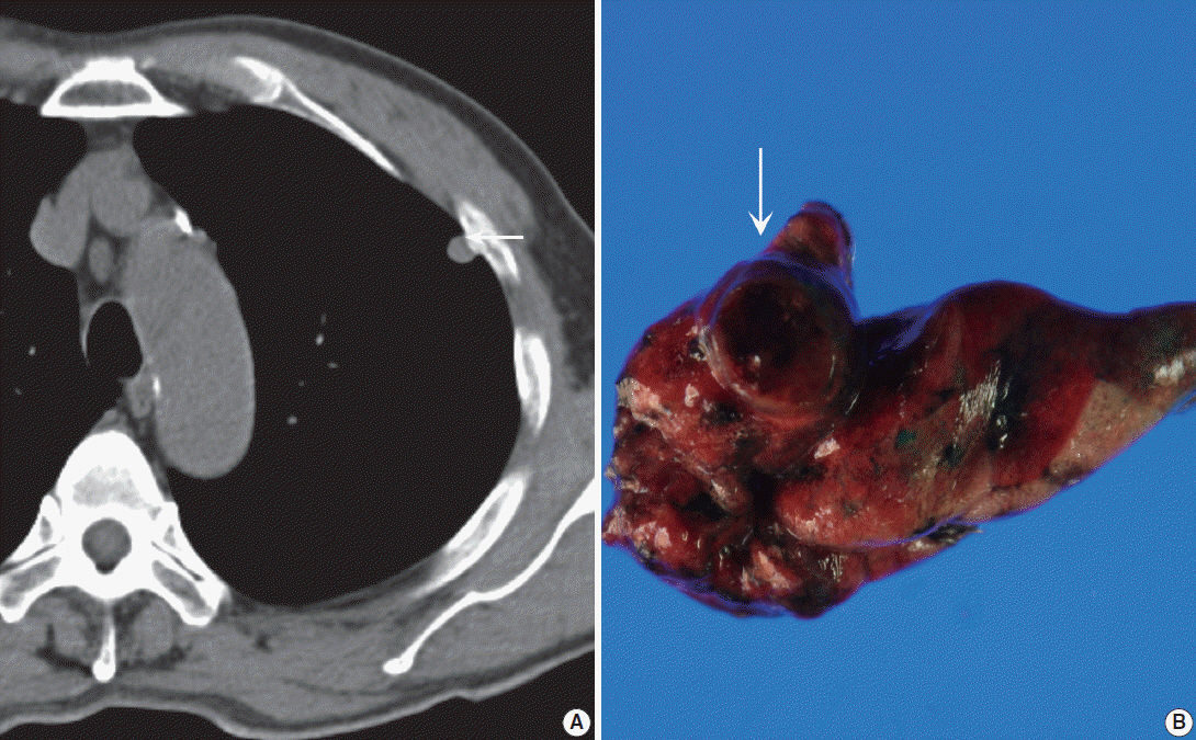
Fig. 1. (A) Precontrast chest computed tomography shows a well-delineated low-attenuated oval mass (arrow) with slightly high density delete the portion in the subpleural area. (B) Gross photo shows a well-demarcated ovoid mass (arrow) with a mucous gelatinous texture with focal hemorrhage.
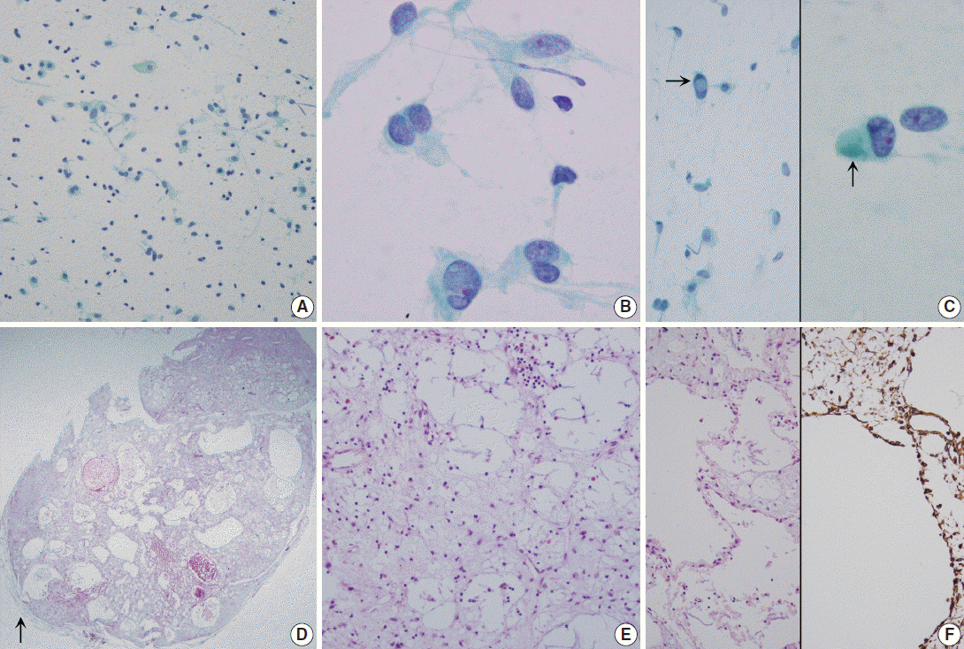
Fig. 2. (A–C) Touch imprint cytology. (A) Hypocellular smear shows many scattered inflammatory cells and macrophages. Note the background amorphous mucin-like metachromatic materials. (B) High-power view shows round to spindle cells having a moderate amount of granular cytoplasm and euchromatic round nuclei. (C) Note rare intranuclear inclusions (left, arrow) and cytoplasmic globular materials (right, arrow) (A–C, Papanicolaou stain). (D–F) Histologic findings. (D) Low-power view shows a well-demarcated bluish mass, and the mass is composed of predominantly chondromyxoid stroma. Arrow indicates normal pulmonary parenchyma. (E) High-power view shows that the mass is composed of abundant bluish chondromyxoid stroma and the paucicellular elements including scattered spindle cells, intermixed macrophages, mature lymphocytes, and some eosinophils. (F) Microcysts are surrounded by attenuated flattened cells or no lining (left, H&E stain; right, Vimentin immunostain).

Fig. 3. Ultrastructural examination reveals spindle to round cells with round euchromatic nuclei and cytoplasm having many lysosomes, mitochondria, and rough endoplasmic reticulums. Extracellular amorphous mucin material (arrow) is observed (left, ×2,500). Multiple ruffled cell surfaces (thin arrow) and abundant collagen fibrils (thick arrow) are also observed (right, ×3,500).
Reference
-
1. Meir K, Maly A, Doviner V, Maly B. Intraoperative cytologic diagnosis of unsuspected cardiac myxoma: a case report. Acta Cytol. 2004; 48:565–8.2. Alaiti S, Nelson FP, Ryoo JW. Solitary cutaneous myxoma. J Am Acad Dermatol. 2000; 43(2 Pt 2):377–9.
Article3. Nadrous HF, Krowka MJ, Myers JL, Allen MS, Sabri AN. Tracheal myxoma: a rare benign tracheal tumor. Mayo Clin Proc. 2004; 79:931–3.
Article4. Özdoğan S, Fidan A, Sarac¸ G, Çağlayan B, Uçmakli E. Concomitant occurrence of lung adenocarcinoma and endobronchial myxoma: a case report. Turk Respir J. 2004; 5:124–7.5. Shilo K, Miettinen M, Travis WD, Timens W, Nogueira R, Franks TJ. Pulmonary microcystic fibromyxoma: report of 3 cases. Am J Surg Pathol. 2006; 30:1432–5.
Article6. Jin MS, Ha HJ, Baek HJ, Lee JC, Koh JS. Adenomyomatous hamartoma of lung mimicking benign mucinous tumor in fine needle aspiration biopsy: a case report. Acta Cytol. 2008; 52:357–60.7. Handa U, Singhal N, Punia RS, Garg S, Mohan H. Cytologic features and differential diagnosis in a case of extraskeletal mesenchymal chondrosarcoma: a case report. Acta Cytol. 2009; 53:704–6.8. Insabato L, Terracciano LM, Boscaino A, Mozzi RA, Angrisani P, Pettinato G. Extraskeletal myxoid chondrosarcoma with intranuclear vacuoles and microtubular aggregates in the rough endoplasmic reticulum: report of a case with fine needle aspiration and electron microscopy. Acta Cytol. 1990; 34:858–62.9. Munjal K, Pancholi V, Rege J, Munjal S, Bhandari V, Nahar R. Fine needle aspiration cytology in mediastinal myxoid liposarcoma: a case report. Acta Cytol. 2007; 51:456–8.10. Orlandi A, Ciucci A, Ferlosio A, Pellegrino A, Chiariello L, Spagnoli LG. Increased expression and activity of matrix metalloproteinases characterize embolic cardiac myxomas. Am J Pathol. 2005; 166:1619–28.
Article
- Full Text Links
-
- Actions
-
Cited
- CITED
-
- Close
- Share
-
- Similar articles
-
- A Case of Multiple Right Atrial Myxomas with Pulmonary Embolism
- A Case of Left Atrial Myxoma Presenting as Acute Pulmonary Edema
- Successful Surgical Treatment of a Right Atrial Myxoma Complicated by Pulmonary Embolism
- Left Ventricular Myxoma Associated Acute Pulmonary Embolism
- Case of Left Atrium Myxoma with Inferior Vena Caval Thrombus and Pulmonary Embolism Complicated with Budd-Chiari Syndrome
