Esophageal Lymphoepithelioma-Like Carcinoma with Unique Daisy-Like Appearance
- Affiliations
-
- 1Department of Gastroenterology, Yuzuncu Yil University Medical Faculty, Van, Turkey. drsehmusolmez@gmail.com
- 2Department of Medical Oncology, Yuzuncu Yil University Medical Faculty, Van, Turkey.
- 3Department of Radiology, Yuzuncu Yil University Medical Faculty, Van, Turkey.
- 4Department of Surgery, Yuzuncu Yil University Medical Faculty, Van, Turkey.
- 5Department of Pathology, Yuzuncu Yil University Medical Faculty, Van, Turkey.
- KMID: 2380414
- DOI: http://doi.org/10.5946/ce.2015.48.6.549
Abstract
- Due to differences in prognosis and management, it is important to subclassify esophageal carcinoma. Esophageal lymphoepithelioma-like carcinoma (LELC) is extremely rare, with only a few cases reported to date. Review of the literature revealed case reports describing lesions with similar histology. We present a 69-year-old man with a giant pedunculated-polypoid lesion of the esophagus shrinking the lumen. Endoscopic excision of the tumor was performed and final histopathological diagnosis was confirmed to be LELC. In contrast to a previous case with a more aggressive course and a recurrent lesion, our patient died of his disease within 8 months of diagnosis. Here we discuss the endoscopic and radiologic findings of the case and a review of the literature.
Figure
-

Fig. 1. Thoracic computed tomography (CT) scan with intravenous contrast medium administration. (A) Examination revealed nodular soft tissue lesion narrowing the esophageal lumen at the level of the carina. The size of the lesion measured 13×10 mm in diameter. (B) Enlargement of the lesion was detected and mediastinal lymphadenopathies were established at 6-month follow-up on thoracic CT scan. (C) Abdominal CT imaging revealed liver metastasis.
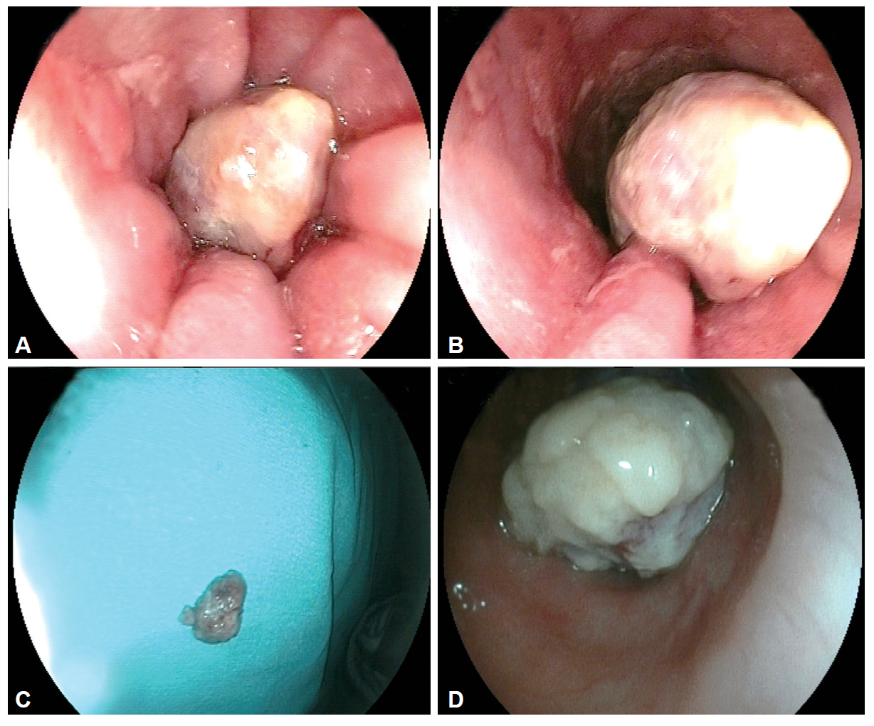
Fig. 2. Upper endoscopic examination. (A) Endoscopy revealed a giant pedunculated polypoid lesion partially shrinking the esophageal lumen at the level of 23 cm from the incisor. Yellowish-white exudative secretion over the polypoid lesion was determined. The combination of polypoid tumor and mucosal changes constituted the unique endoscopic “daisy-like” appearance. (B) Esophageal mucosa was irregular, reddish colored, and contained erosion from the level of the lesion to the cardioesophageal junction. (C) Endoscopic excision polypectomy was performed with snare. (D) Endoscopic examination revealed a giant pedunculated-polypoid lesion totally obliterating the esophageal lumen at the level of 24 cm from the incisor. The lesion surface was ulcerative and had a white exudative feature.

Fig. 3. Histopathologic examination of the lesion. (A) The tumor is characterized by sheets of undifferentiated cells with large nuclei and poorly defined cytoplasmic borders, imparting a syncytial appearance (H&E stain, ×200). (B) Strong cytoplasmic staining with cytokeratin in lymphoepithelioma-like carcinoma (CK, ×200).
Reference
-
1. Tsang WY, Kuo TT, Chan JK. Lymphoepithelial carcinoma. In : Barnes L, Eveson JW, Reichart P, editors. Pathology and Genetics of Head and Neck Tumours. Lyon: IARC Press;2005. p. 251–252.2. Gurzu S, Szentirmay Z, Bara T, et al. Non-Epstein-Barr virus associated lymphoepithelioma-like carcinoma of the esophagogastric junction with microsatellite instability, K-ras wild type. Pathol Res Pract. 2013; 209:128–131.
Article3. Angulo-Pernett F, Smythe WR. Primary lymphoepithelioma of the esophagus. Ann Thorac Surg. 2003; 76:603–605.
Article4. Kuo T, Hsueh C. Lymphoepithelioma-like salivary gland carcinoma in Taiwan: a clinicopathological study of nine cases demonstrating a strong association with Epstein-Barr virus. Histopathology. 1997; 31:75–82.
Article5. Tardío JC, Cristóbal E, Burgos F, Menárguez J. Absence of EBV genome in lymphoepithelioma-like carcinomas of the larynx. Histopathology. 1997; 30:126–128.
Article6. Regaud C, Reverchon L. Sur uncasd’epitheliome epidermoide developpe dans les massif maxillaire superieur. Rev Laryngol Otol Rhinol. 1921; 42:369–378.7. Schmincke A. Über lymphoepitheliale Geschwülste. Beitr Pathol Anat. 1921; 68:161–170.8. Nakasono M, Hirokawa M, Suzuki M, et al. Lymphoepithelioma-like carcinoma of the esophagus: report of a case with non-progressive behavior. J Gastroenterol Hepatol. 2007; 22:2344–2347.
Article9. Falzarano SM, Mourmouras V, Mastrogiulio MG, La Magra C, Vindigni C. Undifferentiated gastric carcinoma with lymphoid stroma (lymphoepithelioma-like carcinoma/medullary carcinoma). Pathologica. 2009; 101:15–17.10. Papalambros E, Felekouras E, Pikoulis E, et al. Epstein-Barr virus: associated adenocarcinoma of the stomach: a rare entity with distinct characteristics. J BUON. 2003; 8:329–331.11. Sashiyama H, Nozawa A, Kimura M, et al. Case report: a case of lymphoepithelioma-like carcinoma of the oesophagus and review of the literature. J Gastroenterol Hepatol. 1999; 14:534–539.
Article
- Full Text Links
-
- Actions
-
Cited
- CITED
-
- Close
- Share
-
- Similar articles
-
- Lymphoepithelioma-like Carcinoma of the Renal Pelvis
- A Case of Lymphoepithelioma - Like Carcinoma of Uterine Cervix
- A Case of Lymphoepithelioma-Like Carcinoma in the Thyroid Gland
- A case of submucosal gastric lymphoepithelioma-like carcinoma
- A Case of Lymphoepithelioma-like Carcinoma Arising from the Parotid Gland
