Advanced Imaging Technology Other than Narrow Band Imaging
- Affiliations
-
- 1Digestive Disease Center, Soonchunhyang University Hospital, Soonchunhyang University College of Medicine, Seoul, Korea. chojhmd@naver.com
- KMID: 2380407
- DOI: http://doi.org/10.5946/ce.2015.48.6.503
Abstract
- To improve the detection rate of gastrointestinal tumors, image-enhanced endoscopy has been widely used during screening and surveillance endoscopy in Korea. In addition to narrow band imaging (NBI) with/without magnification, various types of electronic chromoendoscopies have been used, including autofluorescence imaging, I-scan, and flexible spectral imaging color enhancement. These technologies enable the accurate characterization of tumors because they enable visualization of microvascular and microsurface patterns. The present review focuses on understanding the principle and clinical applications of advanced imaging technologies other than NBI.
Keyword
Figure
-
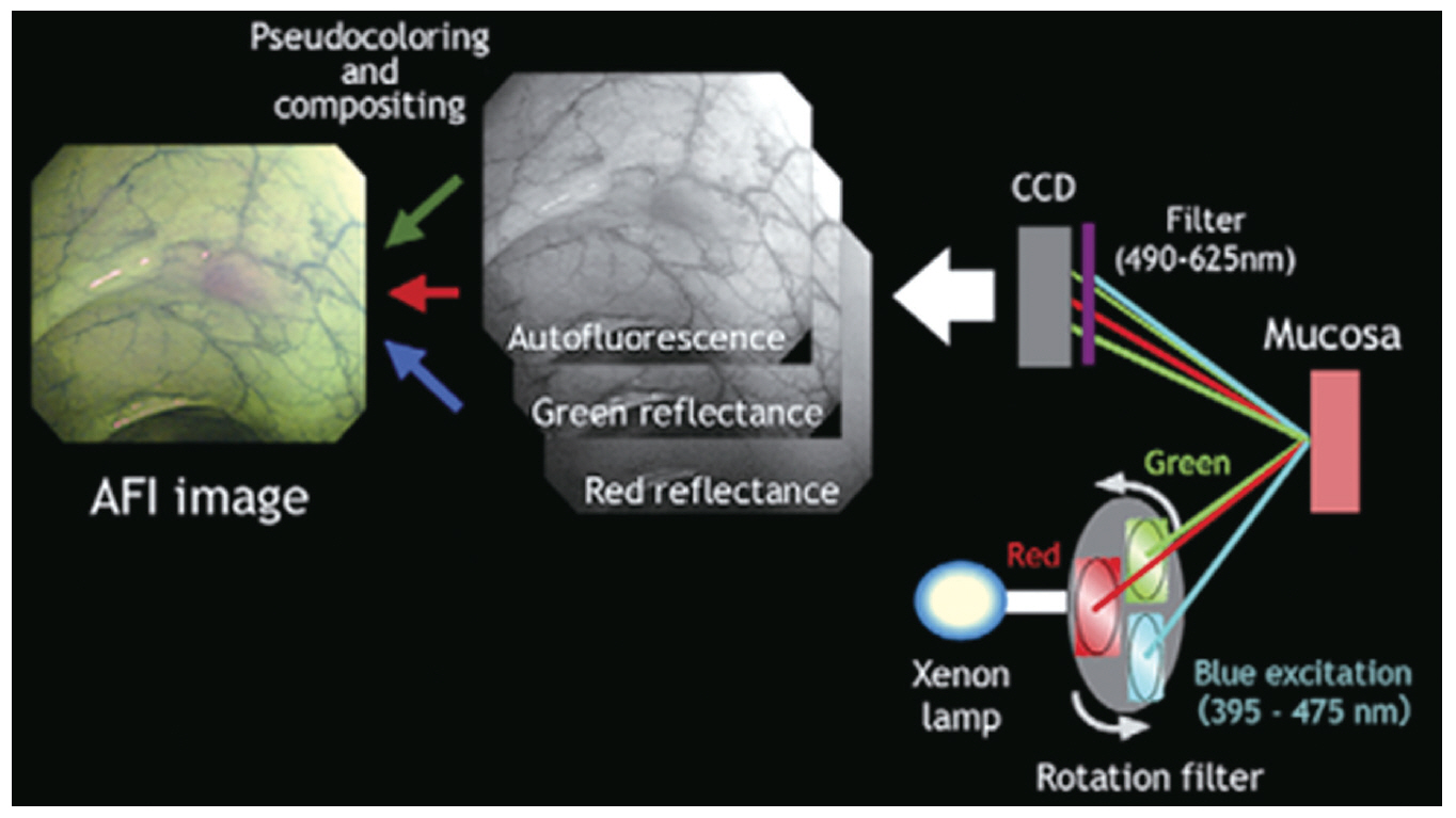
Fig. 1. Diagram of the autofluorescence imaging (AFI) system. Adapted from Uedo et al. [3] with permission from Elsevier. CCD, charge-coupled device.
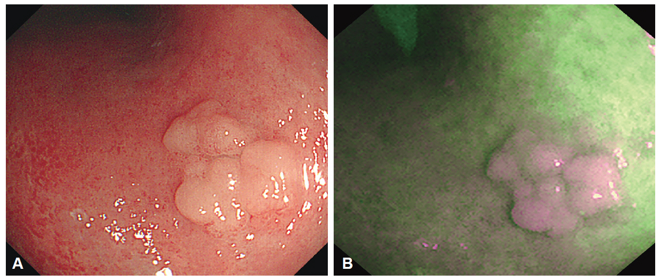
Fig. 2. Endoscopic images of tubular adenoma. (A) A conventional white light image showing an elevated tumor with surface nodularity. (B) An autofluorescence image showing the tumor as a purple area in a green background.
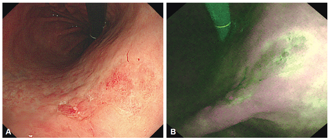
Fig. 3. Endoscopic images of undifferentiated-type early gastric cancer. (A) A conventional white light image. Irregular reddish mucosa observed at the gastric angle. However, tumor margin cannot be differentiated because of similar color of the background mucosa. (B) An autofluorescence image. The tumor appears as purple nodules with a well-defined, green-colored margin.
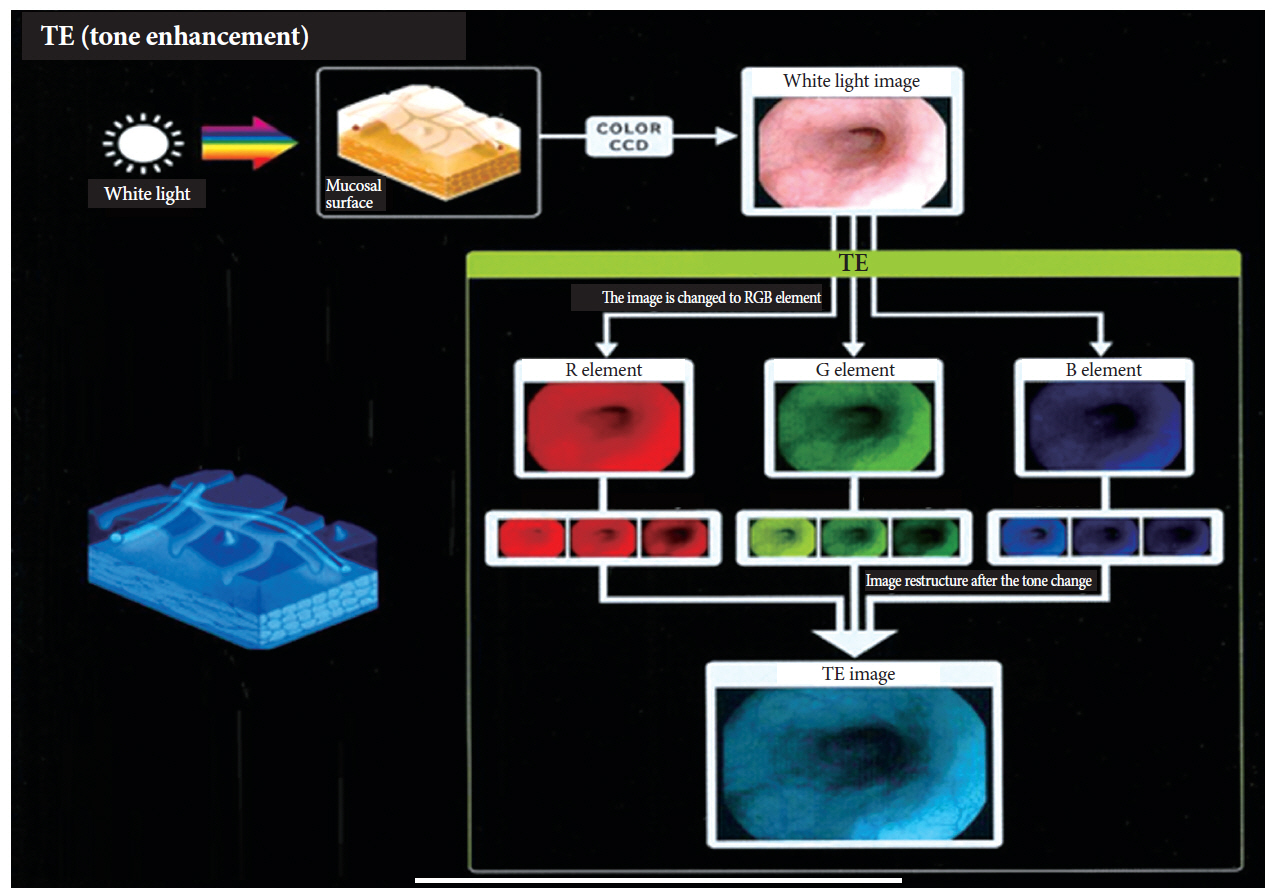
Fig. 4. Principle of tone enhancement by the post-image acquisition software in the I-scan endoscopy system (Courtesy of Pentax, Tokyo, Japan).
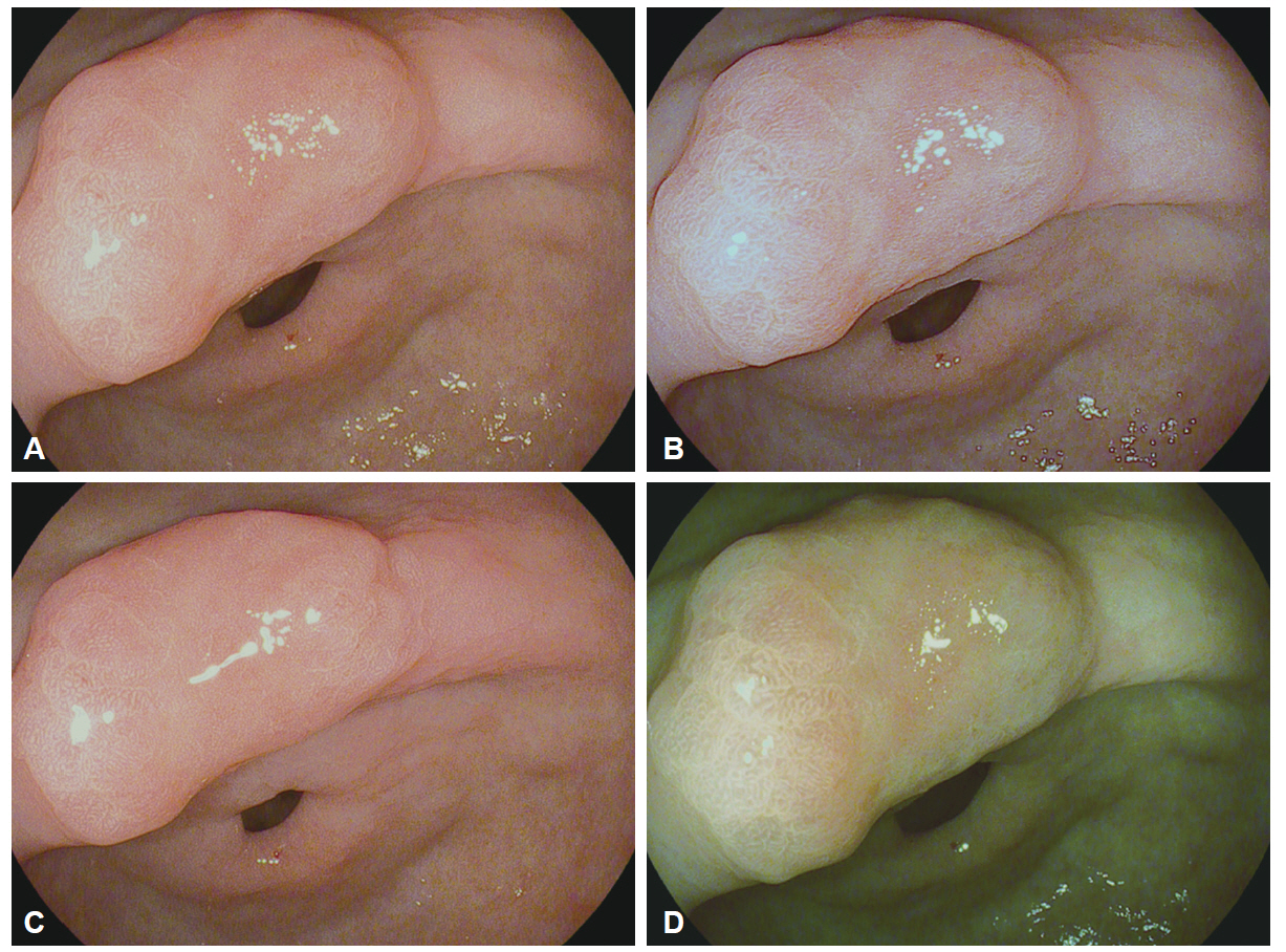
Fig. 5. Endoscopic images of a gastric adenoma. (A) A conventional white light image. (B) I-scan with surface enhancement. (C) I-scan with contrast enhancement. (D) I-scan with tone enhancement.
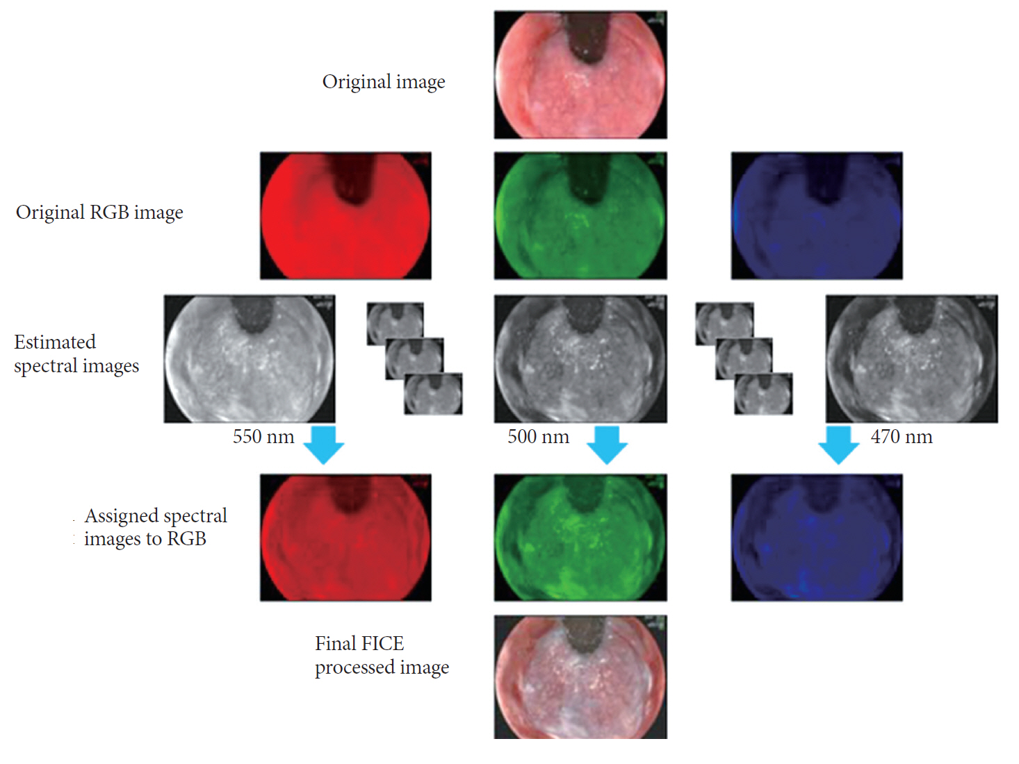
Fig. 6. Principle of the flexible spectral imaging color enhancement (FICE) system (Courtesy of Fujinon Corp., Tokyo, Japan). RGB, red, green, blue.
Reference
-
1. Subramanian V, Ragunath K. Advanced endoscopic imaging: a review of commercially available technologies. Clin Gastroenterol Hepatol. 2014; 12:368–376.e1.
Article2. ASGE Technology Committee, Song LM, Banerjee S, et al. Autofluorescence imaging. Gastrointest Endosc. 2011; 73:647–650.3. Uedo N, Iishi H, Tatsuta M, et al. A novel videoendoscopy system by using autofluorescence and reflectance imaging for diagnosis of esophagogastric cancers. Gastrointest Endosc. 2005; 62:521–528.
Article4. Yoshida Y, Goda K, Tajiri H, Urashima M, Yoshimura N, Kato T. Assessment of novel endoscopic techniques for visualizing superficial esophageal squamous cell carcinoma: autofluorescence and narrow-band imaging. Dis Esophagus. 2009; 22:439–446.
Article5. Suzuki H, Saito Y, Ikehara H, Oda I. Evaluation of visualization of squamous cell carcinoma of esophagus and pharynx using an autofluorescence imaging videoendoscope system. J Gastroenterol Hepatol. 2009; 24:1834–1839.
Article6. Kara MA, Peters FP, Ten Kate FJ, Van Deventer SJ, Fockens P, Bergman JJ. Endoscopic video autofluorescence imaging may improve the detection of early neoplasia in patients with Barrett’s esophagus. Gastrointest Endosc. 2005; 61:679–685.
Article7. Kara MA, Peters FP, Fockens P, ten Kate FJ, Bergman JJ. Endoscopic video-autofluorescence imaging followed by narrow band imaging for detecting early neoplasia in Barrett’s esophagus. Gastrointest Endosc. 2006; 64:176–185.
Article8. Curvers WL, Singh R, Song LM, et al. Endoscopic tri-modal imaging for detection of early neoplasia in Barrett’s oesophagus: a multi-centre feasibility study using high-resolution endoscopy, autofluorescence imaging and narrow band imaging incorporated in one endoscopy system. Gut. 2008; 57:167–172.
Article9. Curvers WL, Singh R, Wallace MB, et al. Identification of predictive factors for early neoplasia in Barrett’s esophagus after autofluorescence imaging: a stepwise multicenter structured assessment. Gastrointest Endosc. 2009; 70:9–17.
Article10. Inoue T, Uedo N, Ishihara R, et al. Autofluorescence imaging videoendoscopy in the diagnosis of chronic atrophic fundal gastritis. J Gastroenterol. 2010; 45:45–51.
Article11. Hanaoka N, Uedo N, Shiotani A, et al. Autofluorescence imaging for predicting development of metachronous gastric cancer after Helicobacter pylori eradication. J Gastroenterol Hepatol. 2010; 25:1844–1849.
Article12. Kato M, Kaise M, Yonezawa J, Yoshida Y, Tajiri H. Autofluorescence endoscopy versus conventional white light endoscopy for the detection of superficial gastric neoplasia: a prospective comparative study. Endoscopy. 2007; 39:937–941.
Article13. Kato M, Kaise M, Yonezawa J, et al. Trimodal imaging endoscopy may improve diagnostic accuracy of early gastric neoplasia: a feasibility study. Gastrointest Endosc. 2009; 70:899–906.
Article14. Nakamura M, Tahara T, Shibata T, et al. Diagnostic efficacy of autofluorescence and reflectance imaging endoscopy for lateral extension of early gastric cancers. Gastrointest Endosc. 2009; 70:599.
Article15. Otani A, Amano Y, Koshino K, et al. Is autofluorescence imaging endoscopy useful for determining the depth of invasion in gastric cancer? Digestion. 2010; 81:96–103.
Article16. Takeuchi Y, Hanaoka N, Hanafusa M, et al. Autofluorescence imaging of early colorectal cancer. J Biophotonics. 2011; 4:490–497.
Article17. Matsuda T, Saito Y, Fu KI, et al. Does autofluorescence imaging videoendoscopy system improve the colonoscopic polyp detection rate? A pilot study. Am J Gastroenterol. 2008; 103:1926–1932.18. Takeuchi Y, Inoue T, Hanaoka N, et al. Autofluorescence imaging with a transparent hood for detection of colorectal neoplasms: a prospective, randomized trial. Gastrointest Endosc. 2010; 72:1006–1013.
Article19. Ramsoekh D, Haringsma J, Poley JW, et al. A back-to-back comparison of white light video endoscopy with autofluorescence endoscopy for adenoma detection in high-risk subjects. Gut. 2010; 59:785–793.
Article20. Fujiya M, Kohgo Y. Image-enhanced endoscopy for the diagnosis of colon neoplasms. Gastrointest Endosc. 2013; 77:111–118.e5.
Article21. van den Broek FJ, Fockens P, Van Eeden S, et al. Clinical evaluation of endoscopic trimodal imaging for the detection and differentiation of colonic polyps. Clin Gastroenterol Hepatol. 2009; 7:288–295.
Article22. Kuiper T, van den Broek FJ, Naber AH, et al. Endoscopic trimodal imaging detects colonic neoplasia as well as standard video endoscopy. Gastroenterology. 2011; 140:1887–1894.
Article23. Kodashima S, Fujishiro M. Novel image-enhanced endoscopy with i-scan technology. World J Gastroenterol. 2010; 16:1043–1049.
Article24. Neumann H, Fujishiro M, Wilcox CM, Mönkemüller K. Present and future perspectives of virtual chromoendoscopy with i-scan and optical enhancement technology. Dig Endosc. 2014; 26 Suppl 1:43–51.
Article25. Hoffman A, Basting N, Goetz M, et al. High-definition endoscopy with i-Scan and Lugol’s solution for more precise detection of mucosal breaks in patients with reflux symptoms. Endoscopy. 2009; 41:107–112.
Article26. Kang HS, Hong SN, Kim YS, et al. The efficacy of i-SCAN for detecting reflux esophagitis: a prospective randomized controlled trial. Dis Esophagus. 2013; 26:204–211.
Article27. Kim MS, Choi SR, Roh MH, et al. Efficacy of I-scan endoscopy in the diagnosis of gastroesophageal reflux disease with minimal change. Clin Endosc. 2011; 44:27–32.
Article28. Li CQ, Li Y, Zuo XL, et al. Magnified and enhanced computed virtual chromoendoscopy in gastric neoplasia: a feasibility study. World J Gastroenterol. 2013; 19:4221–4227.
Article29. Hoffman A, Kagel C, Goetz M, et al. Recognition and characterization of small colonic neoplasia with high-definition colonoscopy using i-Scan is as precise as chromoendoscopy. Dig Liver Dis. 2010; 42:45–50.
Article30. Hoffman A, Sar F, Goetz M, et al. High definition colonoscopy combined with i-Scan is superior in the detection of colorectal neoplasias compared with standard video colonoscopy: a prospective randomized controlled trial. Endoscopy. 2010; 42:827–833.
Article31. Hong SN, Choe WH, Lee JH, et al. Prospective, randomized, back-to-back trial evaluating the usefulness of i-SCAN in screening colonoscopy. Gastrointest Endosc. 2012; 75:1011–1021.e2.
Article32. Neumann H, Vieth M, Fry LC, et al. Learning curve of virtual chromoendoscopy for the prediction of hyperplastic and adenomatous colorectal lesions: a prospective 2-center study. Gastrointest Endosc. 2013; 78:115–120.
Article33. Masci E, Mangiavillano B, Crosta C, et al. Interobserver agreement among endoscopists on evaluation of polypoid colorectal lesions visualized with the Pentax i-Scan technique. Dig Liver Dis. 2013; 45:207–210.
Article34. Lee CK, Lee SH, Hwangbo Y. Narrow-band imaging versus I-Scan for the real-time histological prediction of diminutive colonic polyps: a prospective comparative study by using the simple unified endoscopic classification. Gastrointest Endosc. 2011; 74:603–609.
Article35. Neumann H, Vieth M, Günther C, et al. Virtual chromoendoscopy for prediction of severity and disease extent in patients with inflammatory bowel disease: a randomized controlled study. Inflamm Bowel Dis. 2013; 19:1935–1942.36. Osawa H, Yamamoto H, Yamada N, et al. Diagnosis of endoscopic Barrett’s esophagus by transnasal flexible spectral imaging color enhancement. J Gastroenterol. 2009; 44:1125–1132.
Article37. Osawa H, Yoshizawa M, Yamamoto H, et al. Optimal band imaging system can facilitate detection of changes in depressed-type early gastric cancer. Gastrointest Endosc. 2008; 67:226–234.
Article38. Yoshizawa M, Osawa H, Yamamoto H, et al. Diagnosis of elevated-type early gastric cancers by the optimal band imaging system. Gastrointest Endosc. 2009; 69:19–28.
Article39. Osawa H, Yamamoto H, Miura Y, et al. Diagnosis of extent of early gastric cancer using flexible spectral imaging color enhancement. World J Gastrointest Endosc. 2012; 4:356–361.
Article40. Jung SW, Lim KS, Lim JU, et al. Flexible spectral imaging color enhancement (FICE) is useful to discriminate among non-neoplastic lesion, adenoma, and cancer of stomach. Dig Dis Sci. 2011; 56:2879–2886.
Article41. Chung SJ, Kim D, Song JH, et al. Efficacy of computed virtual chromoendoscopy on colorectal cancer screening: a prospective, randomized, back-to-back trial of Fuji Intelligent Color Enhancement versus conventional colonoscopy to compare adenoma miss rates. Gastrointest Endosc. 2010; 72:136–142.
Article42. Chung SJ, Kim D, Song JH, et al. Comparison of detection and miss rates of narrow band imaging, flexible spectral imaging chromoendoscopy and white light at screening colonoscopy: a randomised controlled back-to-back study. Gut. 2014; 63:785–791.
Article43. Kim YS, Kim D, Chung SJ, et al. Differentiating small polyp histologies using real-time screening colonoscopy with Fuji Intelligent Color Enhancement. Clin Gastroenterol Hepatol. 2011; 9:744–749.e1.
Article44. Pohl J, Lotterer E, Balzer C, et al. Computed virtual chromoendoscopy versus standard colonoscopy with targeted indigocarmine chromoscopy: a randomised multicentre trial. Gut. 2009; 58:73–78.
Article45. Pohl J, Nguyen-Tat M, Pech O, May A, Rabenstein T, Ell C. Computed virtual chromoendoscopy for classification of small colorectal lesions: a prospective comparative study. Am J Gastroenterol. 2008; 103:562–569.
Article46. Kiesslich R, Goetz M, Hoffman A, Galle PR. New imaging techniques and opportunities in endoscopy. Nat Rev Gastroenterol Hepatol. 2011; 8:547–553.
Article47. Isenberg G, Sivak MV Jr, Chak A, et al. Accuracy of endoscopic optical coherence tomography in the detection of dysplasia in Barrett’s esophagus: a prospective, double-blinded study. Gastrointest Endosc. 2005; 62:825–831.
Article48. Arvanitakis M, Hookey L, Tessier G, et al. Intraductal optical coherence tomography during endoscopic retrograde cholangiopancreatography for investigation of biliary strictures. Endoscopy. 2009; 41:696–701.
Article49. Neumann H, Fuchs FS, Vieth M, et al. Review article: in vivo imaging by endocytoscopy. Aliment Pharmacol Ther. 2011; 33:1183–1193.
Article50. Osawa H, Yamamoto H. Present and future status of flexible spectral imaging color enhancement and blue laser imaging technology. Dig Endosc. 2014; 26 Suppl 1:105–115.
Article51. Yoshida N, Hisabe T, Inada Y, et al. The ability of a novel blue laser imaging system for the diagnosis of invasion depth of colorectal neoplasms. J Gastroenterol. 2014; 49:73–80.
Article52. Yoshida N, Yagi N, Inada Y, et al. Ability of a novel blue laser imaging system for the diagnosis of colorectal polyps. Dig Endosc. 2014; 26:250–258.
Article
- Full Text Links
-
- Actions
-
Cited
- CITED
-
- Close
- Share
-
- Similar articles
-
- Advanced Imaging Technology in Biliary Tract Diseases:Narrow-Band Imaging of the Bile Duct
- Usefulness of Narrow-Band Imaging in Endoscopic Submucosal Dissection of the Stomach
- Narrow Band Imaging: Technology Basis and Research and Development History
- Endoscopic Imaging in Barrett's Oesophagus: Applications in Routine Clinical Practice and Future Outlook
- Interobserver Agreement in Using Magnifying Narrow Band Imaging System
