Korean J Ophthalmol.
2017 Jun;31(3):257-262. 10.3341/kjo.2016.0021.
Measurement of Angle Kappa Using Ultrasound Biomicroscopy and Corneal Topography
- Affiliations
-
- 1Department of Ophthalmology, Chung-Ang University College of Medicine, Seoul, Korea. lk1246@hanmail.net
- KMID: 2379882
- DOI: http://doi.org/10.3341/kjo.2016.0021
Abstract
- PURPOSE
To introduce a new convenient and accurate method to measure the angle kappa using ultrasound biomicroscopy (UBM) and corneal topography.
METHODS
Data from 42 eyes (13 males and 29 females) were analyzed in this study. The angle kappa was measured using Orbscan II and calculated with UBM and corneal topography. The angle kappa of the dominant eye was compared with measurements by Orbscan II.
RESULTS
The mean patient age was 36.4 ± 13.8 years. The average angle kappa measured by Orbscan II was 3.98°± 1.12°, while the average angle kappa calculated with UBM and corneal topography was 3.19°± 1.15°. The difference in angle kappa measured by the two methods was statistically significant (p < 0.001). The two methods showed good reliability (intraclass correlation coefficient, 0.671; p < 0.001). Bland-Altman plots were used to demonstrate the agreement between the two methods.
CONCLUSIONS
We designed a new method using UBM and corneal topography to calculate the angle kappa. This method is convenient to use and allows for measurement of the angle kappa without an expensive device.
Figure
-
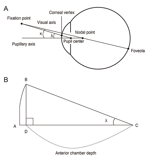
Fig. 1 (A) Schematic representation of the angle kappa components. (B) Schematic diagram showing how to measure angle kappa using ultrasound biomicroscopy and corneal topography. κ = angle kappa; λ = angle lambda; A = point of the cornea that falls on the pupillary axis; B = point of the cornea that lies within the line of sight; C = center of the pupil; D = point of the pupillary axis that meets the vertical line that passes through point B.
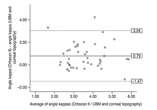
Fig. 2 Bland-Altman plots showing agreement between the two methods. Bold line shows the mean of the difference between the two methods; the dashed line indicates the 95% limit of agreement. UBM = ultrasound biomicroscopy.
Reference
-
1. Park CY, Oh SY, Chuck RS. Measurement of angle kappa and centration in refractive surgery. Curr Opin Ophthalmol. 2012; 23:269–275.2. Basmak H, Sahin A, Yildirim N, et al. Measurement of angle kappa with synoptophore and Orbscan II in a normal population. J Refract Surg. 2007; 23:456–460.3. Emsley HH, editor. Visual optics. 5th ed. London: Hatton Press;1952. p. 523.4. Uozato H, Guyton DL. Centering corneal surgical procedures. Am J Ophthalmol. 1987; 103(3 Pt 1):264–275.5. Sung YJ, Nam SM, Lew H. Measurement of angle lambda using pentacam in normal and exotropic children. J Korean Ophthalmol Soc. 2015; 56:1263–1267.6. Hernandez-Camarena JC, Chirinos-Saldana P, Navas A, et al. Repeatability, reproducibility, and agreement between three different Scheimpflug systems in measuring corneal and anterior segment biometry. J Refract Surg. 2014; 30:616–621.7. Dominguez-Vicent A, Monsalvez-Romin D, Perez-Vives C, et al. Measurement of angle kappa with Orbscan II and Galilei G4: effect of accommodation. Graefes Arch Clin Exp Ophthalmol. 2014; 252:249–255.8. Kim HK, Cho KJ. The angle kappa in dominant and non-dominant eye. J Korean Ophthalmol Soc. 2015; 56:494–498.9. London R, Wick BC. Changes in angle lambda during growth: theory and clinical applications. Am J Optom Physiol Opt. 1982; 59:568–572.10. Le Grand Y, El Hage SG, editors. Physiological optics. New York: Springer-Verlag;1980. p. 73.11. Braaf B, van de Watering TC, Spruijt K, et al. Calculating angle lambda (λ) using zernike tilt measurements in specular reflection corneal topography. J Optom. 2009; 2:207–214.12. Hashemi H, KhabazKhoob M, Yazdani K, et al. Distribution of angle kappa measurements with Orbscan II in a population-based survey. J Refract Surg. 2010; 26:966–971.13. Choi SR, Kim US. The correlation between angle kappa and ocular biometry in Koreans. Korean J Ophthalmol. 2013; 27:421–424.14. Reddy AR, Pande MV, Finn P, El-Gogary H. Comparative estimation of anterior chamber depth by ultrasonography, Orbscan II, and IOLMaster. J Cataract Refract Surg. 2004; 30:1268–1271.15. Al Farhan HM. Agreement between Orbscan II, VuMAX UBM and Artemis-2 very-high frequency ultrasound scanner for measurement of anterior chamber depth. BMC Ophthalmol. 2014; 14:20.16. Vetrugno M, Cardascia N, Cardia L. Anterior chamber depth measured by two methods in myopic and hyperopic phakic IOL implant. Br J Ophthalmol. 2000; 84:1113–1116.17. Rozema JJ, Wouters K, Mathysen DG, Tassignon MJ. Overview of the repeatability, reproducibility, and agreement of the biometry values provided by various ophthalmic devices. Am J Ophthalmol. 2014; 158:1111–1120.e1.
- Full Text Links
-
- Actions
-
Cited
- CITED
-
- Close
- Share
-
- Similar articles
-
- Quantified Values of Anterior Chamber Depth and Angle Measurements Using Ultrasound Biomicroscopy and Topography
- The Angle Kappa in Dominant and Non-Dominant Eye
- Ultrasound Biomicroscopy (UBM plus, model P45, Paradigm(R)): Intraobserver Reproducibility and Agreement of Measurements
- Ciliary Sulcus Size according to Refractive Error using Ultrasound Biomicroscopy
- Comparison of Corneal Thickness Measurements Between Orbscan Topography System and Ultrasonic Pachymetry before and after LASIK
