Maxillary molar derotation and distalization by using a nickel-titanium wire fabricated on a setup model
- Affiliations
-
- 1Division of Orthodontics, Department of Dentistry, Ewha Womans University Mokdong Hospital, Seoul, Korea. yschun@ewha.ac.kr
- KMID: 2379559
- DOI: http://doi.org/10.4041/kjod.2017.47.4.268
Abstract
- The purpose of this article is to introduce a simple appliance that uses a setup model and a nickel-titanium (Ni-Ti) wire for correcting the mesial rotation and drift of the permanent maxillary first molar. The technique involves bonding a Ni-Ti wire to the proper position of the target tooth on a setup model, followed by the fabrication of the transfer cap for indirect bonding and its transfer to the patient's teeth. This appliance causes less discomfort and provides better oral hygiene for the patients than do conventional appliances such as the bracket, pendulum, and distal jet. The treatment time is also shorter with the new appliance than with full-fixed appliances. Moreover, the applicability of the new appliance can be expanded to many cases by using screws or splinting with adjacent teeth to improve anchorage.
Keyword
Figure
-
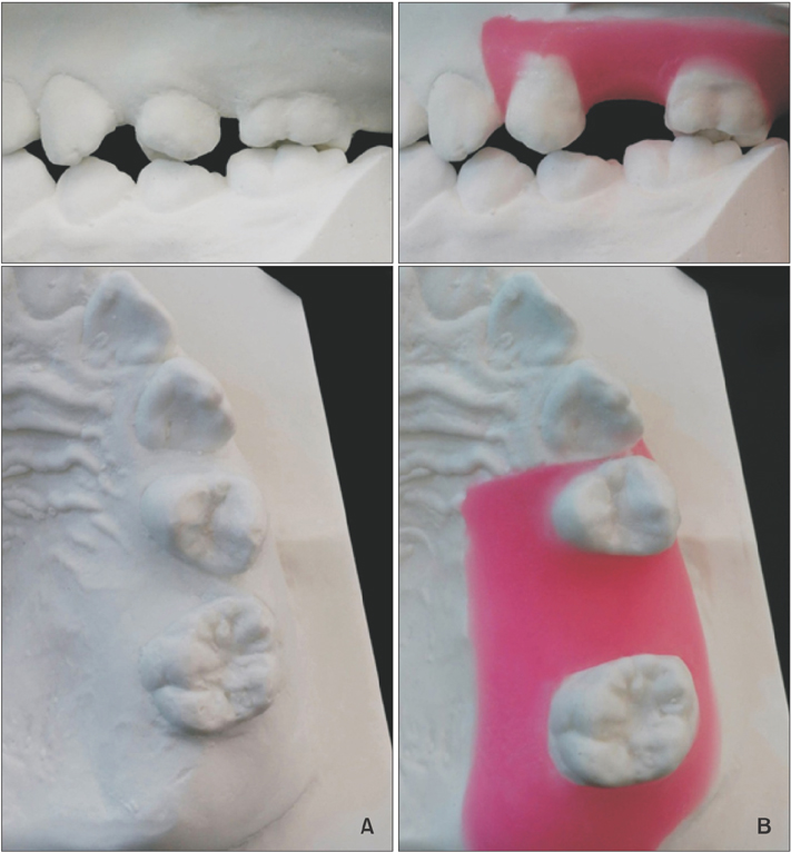
Figure 1 A, Initial model; B, ideal setup model.
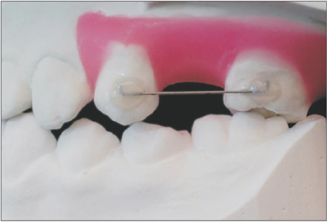
Figure 2 Fabrication of the nickel-titanium (Ni-Ti) spring with a customized resin base. Both ends of the Ni-Ti wire have been bent to prevent unwanted rotation in the resin base.

Figure 3 Fabrication of the transferring resin jig for the nickel-titanium spring with a customized resin base.
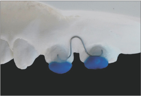
Figure 4 Precise application of the nickel-titanium spring with a customized resin base by transferring the resin jig.
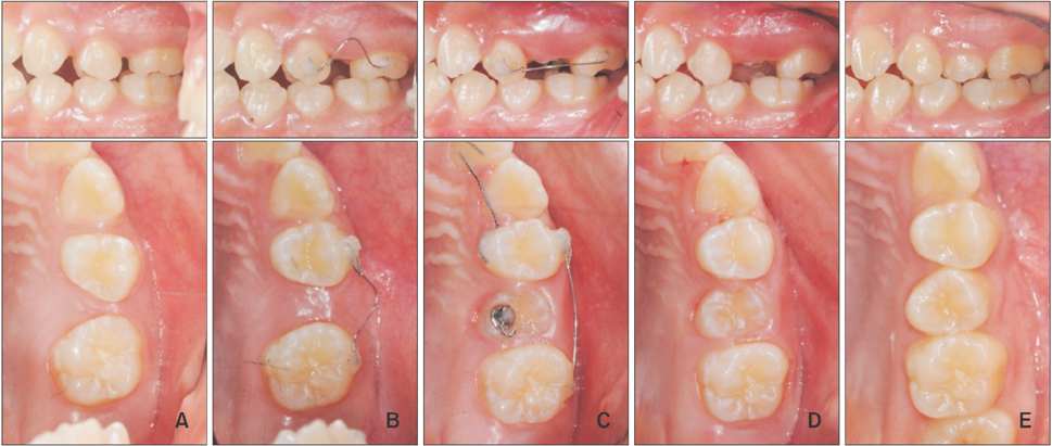
Figure 5 Adequate space is gained through the derotation of the maxillary first premolar and maxillary first molar. A, Pretreatment; B, after 6 weeks; C, after 4 months; D, post-treatment (after 5 months); E, 18 months after treatment. The occlusal button seen in C is bonded for later use during orthodontic traction. However, traction was not applied because the tooth erupted spontaneously.
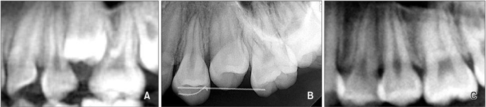
Figure 6 Comparison of the radiographs at A, pretreatment; B, post-treatment; and C, 18 months after treatment.

Figure 7 Three-dimensional superimposition of the pre-treatment (blue) and post-treatment (red) models. The models were scanned using the intraoral scanner (iTero™; Align Technology Inc., Santa Clara, CA, USA), and the models were superimposed using a three-dimensional reverse modeling software (Rapidform 2002; INUS Technology, Seoul, Korea). A, Occlusal view of the maxillary dentition; B, occlusal view of the left maxillary dentition; C, palatal view of the left maxillary dentition; D, buccal view of the left maxillary dentition.
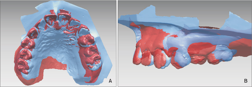
Figure 8 Three-dimensional superimposition of the setup model (blue) and post-treatment (red) model. The maxillary left first molar is more distally tipped in the post-treatment model than in the setup model. A, Occlusal view of the superimposition; B, buccal view of the superimposition.
Cited by 1 articles
-
Treatment of Class I crowding using simple tubes bonded with customized resin coverings: A case report
Seo-Rin Jeong, Hye-In Kim, Sung-Hoon Lim
Korean J Orthod. 2019;49(2):116-123. doi: 10.4041/kjod.2019.49.2.116.
Reference
-
1. Dahlquist A, Gebauer U, Ingervall B. The effect of a transpalatal arch for the correction of first molar rotation. Eur J Orthod. 1996; 18:257–267.
Article2. Bondemark L, Kurol J. Distalization of maxillary first and second molars simultaneously with repelling magnets. Eur J Orthod. 1992; 14:264–272.
Article3. Fontana M, Cozzani M, Caprioglio A. Non-compliance maxillary molar distalizing appliances: an overview of the last decade. Prog Orthod. 2012; 13:173–184.
Article4. Gurgel Jde A, Pinzan-Vercelino CR, Bramante FS, Rivera AP. Distalization of maxillary molars using a lever arm and mini-implant. Orthodontics (Chic.). 2013; 14:e140–e149.5. Hourfar J, Ludwig B, Kanavakis G. An active, skeletally anchored transpalatal appliance for derotation, distalization and vertical control of maxillary first molars. J Orthod. 2014; 41:Suppl 1. S24–S32.
Article6. Chiu PP, McNamara JA Jr, Franchi L. A comparison of two intraoral molar distalization appliances: distal jet versus pendulum. Am J Orthod Dentofacial Orthop. 2005; 128:353–365.
Article7. Caprioglio A, Cafagna A, Fontana M, Cozzani M. Comparative evaluation of molar distalization therapy using pendulum and distal screw appliances. Korean J Orthod. 2015; 45:171–179.
Article8. Klukowska M, Bader A, Erbe C, Bellamy P, White DJ, Anastasia MK, et al. Plaque levels of patients with fixed orthodontic appliances measured by digital plaque image analysis. Am J Orthod Dentofacial Orthop. 2011; 139:e463–e470.
Article9. Wiechmann D, Gerss J, Stamm T, Hohoff A. Prediction of oral discomfort and dysfunction in lingual orthodontics: a preliminary report. Am J Orthod Dentofacial Orthop. 2008; 133:359–364.
Article10. Locatelli R, Bednar J, Dietz VS, Gianelly AA. Molar distalization with superelastic NiTi wire. J Clin Orthod. 1992; 26:277–279.11. Kim M, Kim M, Chun YS. Molar uprighting by a nickel-titanium spring based on a setup model. Am J Orthod Dentofacial Orthop. 2014; 146:119–123.
Article12. Choi KH, Lee Y, Kim M, Chun YS. Correction of palatally displaced maxillary lateral incisors without brackets. Korean J Orthod. 2013; 43:201–206.
Article
- Full Text Links
-
- Actions
-
Cited
- CITED
-
- Close
- Share
-
- Similar articles
-
- Immediate changes in the mandibular dentition after maxillary molar distalization using headgear
- Zygoma-gear appliance for intraoral upper molar distalization
- Effects of heat treatment on the load-deflection properties of nickel-titanium wire
- Combined treatment with headgear and the Frog appliance for maxillary molar distalization: a randomized controlled trial
- Noncompliance screw supported maxillary molar distalization in a parallel manner
