Differential diagnosis of periapical cyst using collagen birefringence pattern of the cyst wall
- Affiliations
-
- 1Department of Conservative Dentistry, Gangneung-Wonju National University College of Dentistry, Gangneung, Korea. mendo7@gwnu.ac.kr
- 2Department of Oral Pathology, Gangneung-Wonju National University College of Dentistry, Gangneung, Korea. sklee@gwnu.ac.kr
- KMID: 2379460
- DOI: http://doi.org/10.5395/rde.2017.42.2.111
Abstract
OBJECTIVES
Periapical lesions, including periapical cyst (PC), periapical granuloma (PG), and periapical abscess (PA), are frequently affected by chemical/physical damage during root canal treatment or severe bacterial infection, and thus, the differential diagnosis of periapical lesions may be difficult due to the presence of severe inflammatory reaction. The aim of this study was to make differential diagnosis among PC, PG, and PA under polarizing microscope.
MATERIALS AND METHODS
The collagen birefringence patterns of 319 cases of PC (n = 122), PG (n = 158), and PA (n = 39) obtained using a polarizing microscope were compared. In addition, 6 cases of periodontal fibroma (PF) were used as positive controls.
RESULTS
Collagen birefringence was condensed with a thick, linear band-like pattern in PC, but was short and irregularly scattered in PG, and scarce or absent in PA. PF showed intense collagen birefringence with a short, palisading pattern but no continuous band-like pattern. The linear band-like birefringence in PC was ascribed to pre-existing expansile tensile stress of the cyst wall.
CONCLUSIONS
In this study all PCs (n = 122) were distinguishable from PGs and PAs by their characteristic birefringence, despite the absence of lining epithelium (n = 20). Therefore, the authors suggest that the presence of linear band-like collagen birefringence of the cyst wall aids the diagnostic differentiation of PC from PG and PA.
MeSH Terms
Figure
-
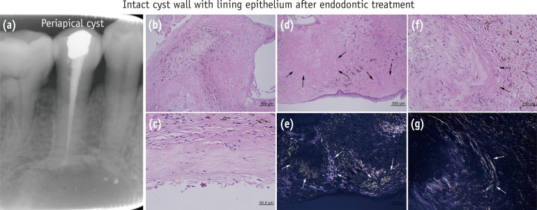
Figure 1 Periapical cyst with lining epithelium after endodontic treatment, noted the linear band-like birefringence in collagen bundles (arrows) encircling the cyst wall. (e) and (g), polarized views of (d) and (f), respectively.
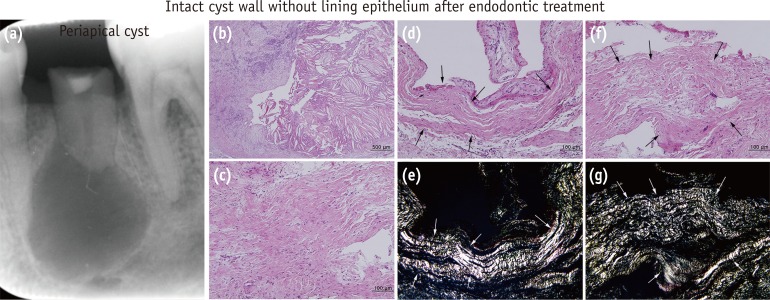
Figure 2 Periapical cyst with no lining epithelium after endodontic treatment, noted strong linear band-like birefringence parallel to the cyst wall in collagen bundles (arrows). (e) and (g), polarized views of (d) and (f), respectively.
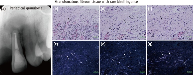
Figure 3 Periapical granuloma, note the weak and rudimentary birefringence in degenerating collagen bundles (arrows) of this granulomatous lesion. (c), (e), and (g), polarized views of (b), (d), and (f), respectively.
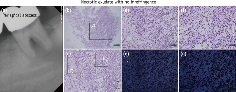
Figure 4 Periapical abscess, note almost no birefringence in the necrotic exudate. (e) and (g), polarized views of (d) and (f), respectively.
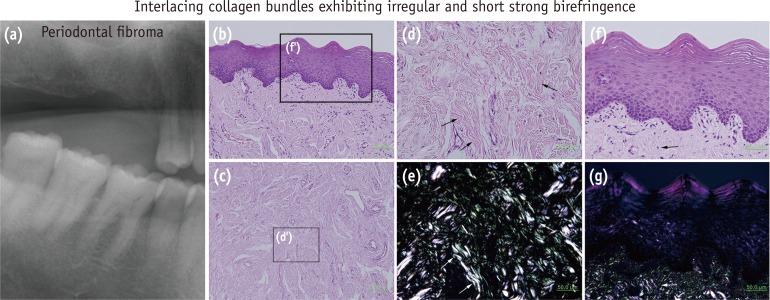
Figure 5 Periodontal fibroma, note strong birefringence with a short and irregularly scattered pattern (arrows). (e) and (g), polarized views of (d) and (f), respectively.
Cited by 1 articles
-
Interplay of collagen and mast cells in periapical granulomas and periapical cysts: a comparative polarizing microscopic and immunohistochemical study
Deepty Bansal, Mala Kamboj, Anjali Narwal, Anju Devi, Nisha Marwah
Restor Dent Endod. 2022;47(1):e12. doi: 10.5395/rde.2022.47.e12.
Reference
-
1. Lin LM, Ricucci D, Lin J, Rosenberg PA. Nonsurgical root canal therapy of large cyst-like inflammatory periapical lesions and inflammatory apical cysts. J Endod. 2009; 35:607–615. PMID: 19410070.
Article2. Valois CR, Costa-Júnior ED. Periapical cyst repair after nonsurgical endodontic therapy-case report. Braz Dent J. 2005; 16:254–258. PMID: 16429195.3. Fernandes M, de Ataide I. Nonsurgical management of periapical lesions. J Conserv Dent. 2010; 13:240–245. PMID: 21217952.
Article4. Moon HI, Kim SH, Hwang YC, Oh BJ, Hwang IN, Kim SH, Jeong SW, Youn C, Oh WM. Pulpal and periapical reaction to formocresol and Depulpin in the rat teeth. J Korean Acad Conserv Dent. 2002; 27:355–362.
Article5. Seltzer S, Bender IB, Smith J, Freedman I, Nazimov H. Endodontic failures - an analysis based on clinical, roentgenographic, and histologic findings. I. Oral Surg Oral Med Oral Pathol. 1967; 23:500–516. PMID: 5227403.6. Nair PN. New perspectives on radicular cysts: do they heal? Int Endod J. 1998; 31:155–160. PMID: 10321160.
Article7. Aggarwal P, Saxena S. Stromal differences in odontogenic cysts of a common histopathogenesis but with different biological behavior: a study with picrosirius red and polarizing microscopy. Indian J Cancer. 2011; 48:211–215. PMID: 21768668.
Article8. Singh HP, Shetty DC, Wadhwan V, Aggarwal P. A quantitative and qualitative comparative analysis of collagen fibers to determine the role of connective tissue stroma on biological behavior of odontogenic cysts: a histochemical study. Natl J Maxillofac Surg. 2012; 3:15–20. PMID: 23251052.
Article9. Vij R, Vij H, Rao NN. Evaluation of collagen in connective tissue walls of odontogenic cysts-a histochemical study. J Oral Pathol Med. 2011; 40:257–262. PMID: 20969631.10. Hirshberg A, Lib M, Kozlovsky A, Kaplan I. The influence of inflammation on the polarization colors of collagen fibers in the wall of odontogenic keratocyst. Oral Oncol. 2007; 43:278–282. PMID: 16919995.
Article11. Martin RB, Lau ST, Mathews PV, Gibson VA, Stover SM. Collagen fiber organization is related to mechanical properties and remodeling in equine bone. A comparison of two methods. J Biomech. 1996; 29:1515–1521. PMID: 8945649.12. Love RM, Firth N. Histopathological profile of surgically removed persistent periapical radiolucent lesions of endodontic origin. Int Endod J. 2009; 42:198–202. PMID: 19228208.
Article13. Garcia CC, Sempere FV, Diago MP, Bowen EM. The post-endodontic periapical lesion: histologic and etiopathogenic aspects. Med Oral Patol Oral Cir Bucal. 2007; 12:E585–E590. PMID: 18059244.14. Martin RB, Ishida J. The relative effects of collagen fiber orientation, porosity, density, and mineralization on bone strength. J Biomech. 1989; 22:419–426. PMID: 2777816.
Article15. Martin RB, Boardman DL. The effects of collagen fiber orientation, porosity, density, and mineralization on bovine cortical bone bending properties. J Biomech. 1993; 26:1047–1054. PMID: 8408087.
Article16. Ganganna K, Shetty P, Shroff SE. Collagen in histologic stages of oral submucous fibrosis: a polarizing microscopic study. J Oral Maxillofac Pathol. 2012; 16:162–166. PMID: 22923884.
Article17. Rowe AJ, Finlay HM, Canham PB. Collagen biomechanics in cerebral arteries and bifurcations assessed by polarizing microscopy. J Vasc Res. 2003; 40:406–415. PMID: 12913333.
Article18. Orberg J, Baer E, Hiltner A. Organization of collagen fibers in the intestine. Connect Tissue Res. 1983; 11:285–297. PMID: 6227451.
Article19. Fackler K, Klein L, Hiltner A. Polarizing light microscopy of intestine and its relationship to mechanical behaviour. J Microsc. 1981; 124:305–311. PMID: 7328641.
Article20. Schultka R, Schmidt T, Hepp WD, Schräpler R, Cech S. Connective tissue of the human fallopian tube mucosa-polarization microscopic detection of collagen using Solaminrot 4B. Acta Histochem. 1989; 86:159–166. PMID: 2514549.21. Shah JS, Jayson MI, Hampson WG. Low tension studies of collagen fibres from ligaments of the human spine. Ann Rheum Dis. 1977; 36:139–145. PMID: 856065.
Article22. Ascenzi A, Bonucci E. Relationship between ultrastructure and ‘pin test’ in osteons. Clin Orthop Relat Res. 1976; 121:275–294.
Article23. Park YS, Cho BH, Lee SH, Shon WJ. Early caries detection using optical coherence tomography: a review of the literature. J Korean Acad Conserv Dent. 2011; 36:367–376.
Article
- Full Text Links
-
- Actions
-
Cited
- CITED
-
- Close
- Share
-
- Similar articles
-
- Anterior stafne bone cyst mimicking periapical cyst: a case report
- Interplay of collagen and mast cells in periapical granulomas and periapical cysts: a comparative polarizing microscopic and immunohistochemical study
- Comparison of digital radiometric featuresbetween radicular cysts and periapical granulomas
- Cutaneous Bronchogenic Cyst of the Anterior Chest Wall
- Immunohistochemical study on the pathological features of odontogenic cyst
