An Unrecognized Foreign Body Retained in the Calcaneus: A Case Report
- Affiliations
-
- 1Department of Radiology, Gangneung Asan Hospital, College of Medicine, University of Ulsan, Gangneung, Korea. sojuch@naver.com
- 2Department of Orthopedic Surgery, Gangneung Asan Hospital, College of Medicine, University of Ulsan, Gangneung, Korea.
- KMID: 2379327
- DOI: http://doi.org/10.3348/jksr.2017.76.6.434
Abstract
- We describe a case of an unrecognized foreign body retained in the calcaneus. The patient denied any history of trauma. The skin overlying the calcaneus was intact with no local signs of inflammation. The retained foreign body was not observed on the radiograph of the calcaneus. Magnetic Resonance Imaging showed a tubular low signal intensity lesion in the calcaneal body, surrounded by strongly enhanced soft tissue and bone marrow edema caused by a foreign body reaction. A foreign body retained in the calcaneus was suspected on the basis of these findings. Surgical exploration and curettage was performed, and a rod shaped wooden fragment was found.
MeSH Terms
Figure
-
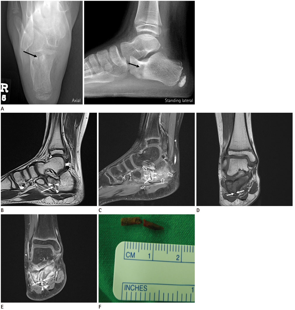
Fig. 1 A 9-year-old boy with an unrecognized foreign body retained in the calcaneus. A. Plain radiographs with axial and standing lateral views of the right foot show a tubular geographic osteolytic lesion in the anterior body of the right calcaneus (arrows) with an ill-defined margin and surrounding sclerotic change. B. Sagittal T2-weighted MR image shows a low signal intensity tubular lesion in the anterior body of the calcaneus (arrow) with surrounding high signal intensity tissue (arrowheads). C. Sagittal fat-saturated enhanced T1-weighted MR image reveals non-enhancement of the tubular lesion, suggesting a foreign body (arrow), whereas the surrounding soft tissue is strongly enhanced (arrowheads), suggestive of a foreign body reaction. Note edematous bone marrow enhancement in the body of the calcaneus (open arrows). D, E. Coronal T1-weighted (D) and fat-saturated enhanced T1 weighted (E) MR images show a cortical defect (arrows) on the plantar surface of the calcaneal body, just below the retained foreign body. Note the focal defect with enhancement in the overlying plantar fascia (arrowheads). F. A gross photograph of the extracted wooden foreign body.
Reference
-
1. Reginato AJ, Ferreiro JL, O'Connor CR, Barbasan C, Arasa J, Bednar J, et al. Clinical and pathologic studies of twenty-six patients with penetrating foreign body injury to the joints, bursae, and tendon sheaths. Arthritis Rheum. 1990; 33:1753–1762.2. Borgia CA. An unusual bone reaction to an organic foreign body in the hand. Clin Orthop Relat Res. 1963; 30:188–193.3. Dürr HR, Stäbler A, Müller PE, Refior HJ. Thorn-induced pseudotumor of the metatarsal. A case report. J Bone Joint Surg Am. 2001; 83-A:580–585.4. Abu Hassan FO. Retained toothpick causing pseudotumor of the first metatarsal: a case report and literature review. Foot Ankle Surg. 2008; 14:32–35.5. Madhar M, Sammous Y, Bouslous J, Messaoudi T, Chafik R, Elhaoury H, et al. Osteitis of the fourth metatarsal caused by a date palm thorn in a child: why the dorsum of the foot is the most commonly injured site. J Foot Ankle Surg. 2013; 52:84–87.6. Guner S, Ceylan MF, Isik D, Guner SI, Ediz L. A case of wooden foreign body retained in the calcaneus. Pak J Med Sci. 2011; 27:932.7. Vidyadhara S, Rao SK. Thorn prick osteomyelitis of the foot in barefoot walkers: a report of four cases. J Orthop Surg (Hong Kong). 2006; 14:222–224.8. Barry M, Maffulli N, Good C. The missed thorn. Acta Orthop Belg. 1992; 58:468–470.9. Dhillon MS, Prasanna HM, Goni V, Nagi ON. Wooden splinter-induced pseudo tumour of the metatarsal. Foot Ankle Surg. 2000; 6:45–48.10. Peterson JJ, Bancroft LW, Kransdorf MJ. Wooden foreign bodies: imaging appearance. AJR Am J Roentgenol. 2002; 178:557–562.
- Full Text Links
-
- Actions
-
Cited
- CITED
-
- Close
- Share
-
- Similar articles
-
- Case Report of Retained Intraorbital Metallic Foreign Body Removal
- A Case of Intracolonic Surgical Sponge misdiagnosed as Intraperitoneal Foreign Body
- Noninfectious Endophthalmitis Caused by an Intraocular Foreign Body Retained for 16 Years
- An Intradiscal Granuloma Due to a Retained Wooden Foreign Body
- Sphenoid sinus foreign body following airbag deployment in the United States: a case report
