Barium Peritonitis Following Upper Gastrointestinal Series: A Case Report
- Affiliations
-
- 1Department of Radiology, Soonchunhyang University College of Medicine, Seoul Hospital, Seoul, Korea. jy0707hwang@schmc.ac.kr
- 2Department of Surgery, Soonchunhyang University College of Medicine, Seoul Hospital, Seoul, Korea.
- KMID: 2379325
- DOI: http://doi.org/10.3348/jksr.2017.76.6.425
Abstract
- We report a rare case of barium peritonitis following an upper gastrointestinal (GI) series and its imaging findings in a 74-year-old female. Barium peritonitis is a rare but life-threatening complication of GI contrast investigation. Therefore, clinical awareness of barium peritonitis as a complication of GI tract contrast investigation would help to prevent such a complication and manage the patients properly.
MeSH Terms
Figure
-
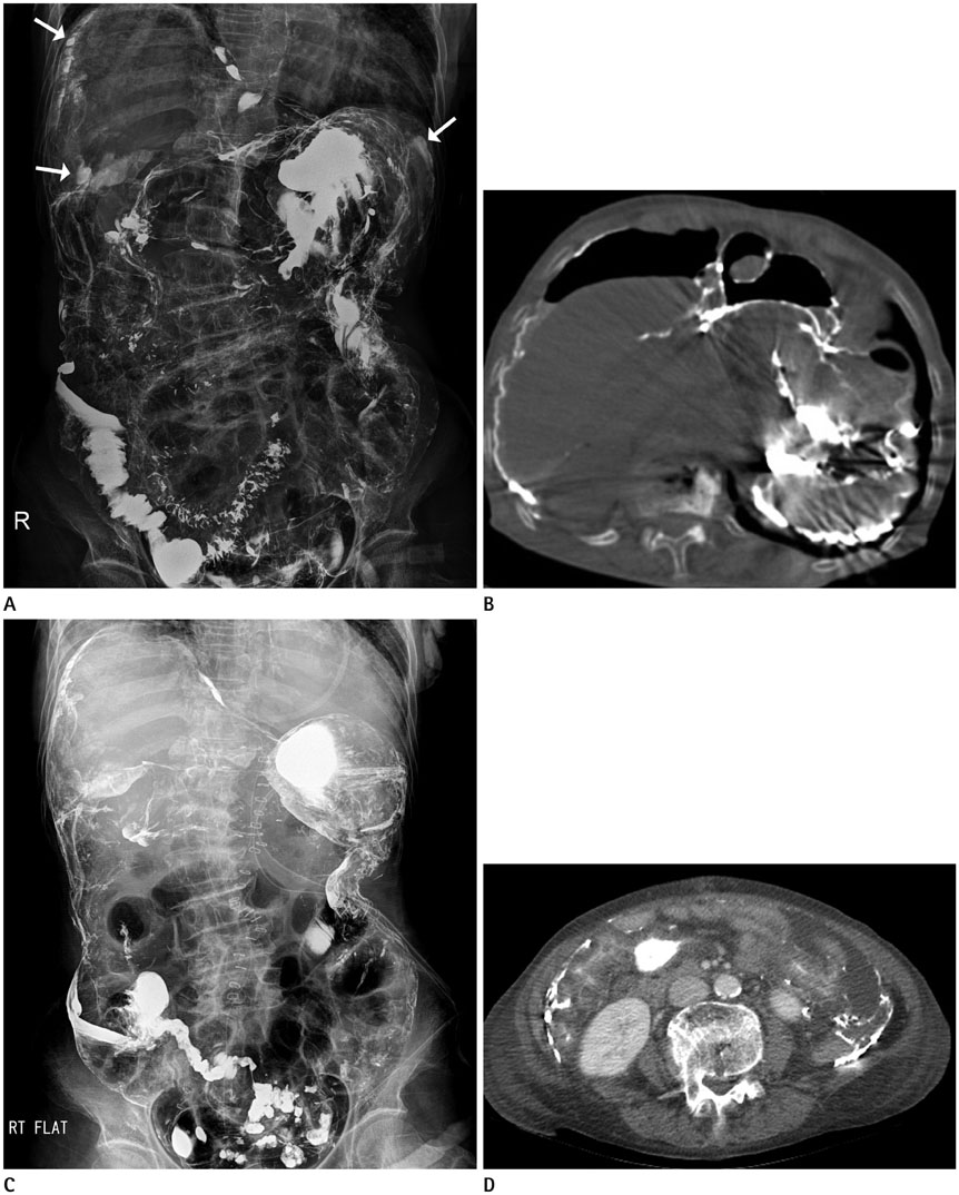
Fig. 1 Pre- and post-operative images of barium peritonitis following an upper gastrointestinal series in a 74-year-old female. A. Pre-operative plain abdominal radiography shows extensive barium spillage in the peritoneal cavity with pneumoperitoneum. Especially, larger amounts of agglomerated barium clumps are seen in bilateral subdiaphragmatic areas (arrows). B. Pre-operative unenhanced axial CT image reveals agglomerated barium clumps adhering to the parietal and visceral surfaces of the peritoneal cavity with a large volume of pneumoperitoneum. Assessment of bowel perforation on current CT images is limited due to unenhanced scan protocol, insufficient bowel distension and beam hardening artifacts. C. Post-operative plain abdominal radiography on the 3rd post-operative day shows residual barium, which outlines the abdominopelvic cavity and probably adheres along the peritoneum. D. Contrast-enhanced axial CT image on the 19th post-operative day shows residual barium and mesenteric fat haziness with ascites. Generalized soft tissue edema is also seen.
Reference
-
1. Himmelmann W. Ueber die perforation im bereich des magen-darmtraktus bei und nach der rontgenbreipassage. Munch Med Wochenschr. 1932; 79:1567–1571.2. Ott DJ, Gelfand DW. Gastrointestinal contrast agents. Indications, uses, and risks. JAMA. 1983; 249:2380–2384.3. Karanikas ID, Kakoulidis DD, Gouvas ZT, Hartley JE, Koundourakis SS. Barium peritonitis: a rare complication of upper gastrointestinal contrast investigation. Postgrad Med J. 1997; 73:297–298.4. Williams SM, Harned RK. Recognition and prevention of barium enema complications. Curr Probl Diagn Radiol. 1991; 20:123–151.5. Noveroske RJ. Intracolonic pressures during barium enema examination. Am J Roentgenol Radium Ther Nucl Med. 1964; 91:852–863.6. Thoeni RF, Margulis AR. Intracolonic pressures during barium-enema studies using the single- and double-contrast techniques. Invest Radiol. 1979; 14:162–165.7. de Feiter PW, Soeters PB, Dejong CH. Rectal perforations after barium enema: a review. Dis Colon Rectum. 2006; 49:261–271.8. Terranova O, Meneghello A, Battocchio F, Martella B, Celi D, Nistri R. Perforations of the extraperitoneal rectum during barium enema. Int Surg. 1989; 74:13–16.9. Zheutlin N, Lasser EC, Rigler LG. Clinical studies on effect of barium in the peritoneal cavity following rupture of the colon. Surgery. 1952; 32:967–979.
- Full Text Links
-
- Actions
-
Cited
- CITED
-
- Close
- Share
-
- Similar articles
-
- An experimental study on barium peritonitis in rats
- Barium Peritonitis due to Inadvertent Vaginal Insertion rather than a Colonic Insertion: 1 Case Report
- Acute Respiratory Failure Caused by Aspiration of High Density Barium: A Case Report
- A Case of Gastrocolic Fistula as a Complication of Colon Cancer
- Gastritis Caused by lngestion of Eggs of Puffer Fish: A Case Report
