Ann Dermatol.
2017 Jun;29(3):349-351. 10.5021/ad.2017.29.3.349.
Multiple Interdigital Nodular Amyloidosis of the Toe: A Unique Presentation of Localized Cutaneous Amyloidosis
- Affiliations
-
- 1Department of Dermatology, The Catholic University of Korea, Uijeongbu St. Mary's Hospital, Uijeongbu, Korea. frankyu123@hotmail.com
- KMID: 2378539
- DOI: http://doi.org/10.5021/ad.2017.29.3.349
Abstract
- No abstract available.
MeSH Terms
Figure
-
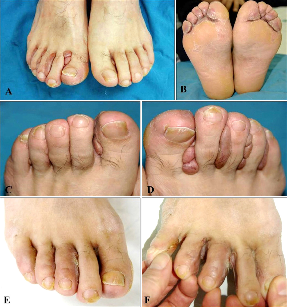
Fig. 1 (A~D) Multiple, slightly erythematous, smooth-surfaced nodules in the interdigital spaces of both feet. (E, F) Interdigital spaces of right foot without recurrence of nodules 6 months after treatment with shave excision.
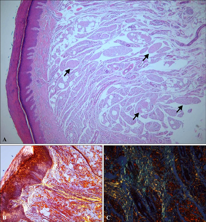
Fig. 2 (A) Diffuse deposition of eosinophilic, amorphous material (arrows) through the entire dermis (H&E, ×40). (B, C) Polarized microscopy image showing birefringent amyloid deposits (Congo red stain under polarized light, B: ×40, C: ×200).
Reference
-
1. Konopinski JC, Seyfer SJ, Robbins KL, Hsu S. A case of nodular cutaneous amyloidosis and review of the literature. Dermatol Online J. 2013; 19:10.
Article2. Ritchie SA, Beachkofsky T, Schreml S, Gaspari A, Hivnor CM. Primary localized cutaneous nodular amyloidosis of the feet: a case report and review of the literature. Cutis. 2014; 93:89–94.3. Borrowman TA, Lutz ME, Walsh JS. Cutaneous nodular amyloidosis masquerading as a foot callus. J Am Acad Dermatol. 2003; 49:307–310.
Article4. Lee DY, Kim YJ, Lee JY, Kim MK, Yoon TY. Primary localized cutaneous nodular amyloidosis following local trauma. Ann Dermatol. 2011; 23:515–518.
Article5. Cornejo KM, Lagana FJ, Deng A. Nodular amyloidosis derived from keratinocytes: an unusual type of primary localized cutaneous nodular amyloidosis. Am J Dermatopathol. 2015; 37:e129–e133.
