Leg Swelling Caused by Heterotopic Ossification Mimicking Deep Vein Thrombosis in a Paraplegic Patient
- Affiliations
-
- 1Department of Neurosurgery, Dongguk University College of Medicine, Dongguk University Ilsan Hospital, Goyang, Korea. ktcho21@naver.com
- 2Department of Rehabilitation Medicine, Dongguk University College of Medicine, Dongguk University Ilsan Hospital, Goyang, Korea.
- KMID: 2378278
- DOI: http://doi.org/10.13004/kjnt.2015.11.2.158
Abstract
- Leg swelling in patients with paraplegia due to spinal cord injury (SCI) occurs for various reasons, including heterotopic ossification (HO), deep vein thrombosis (DVT), fracture, or cellulitis. The clinical presentations of these conditions may overlap in part or in whole and it may occasionally be difficult to distinguish. Of these conditions, DVT and subsequent pulmonary embolism remain significant causes of morbidity and mortality in patients with SCI. Therefore, a prompt diagnostic work-up, particularly for DVT, is essential in patients with SCI, who present with leg swelling. Here, we report a case of leg swelling in a paraplegic patient, resulting from HO mimicking DVT and discuss the differential diagnosis.
MeSH Terms
Figure
-
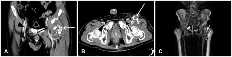
FIGURE 1 Computed tomography venography of the lower extremities. A: Coronal source image of venography shows heterotopic ossification (HO) (white arrow) and narrowing of the left external iliac vein (EIV) (black arrow) compared to the right vein (arrowhead). B: Axial source image of venography shows HO (white arrow) located anteromedial to the femur and displaces the iliopsoas muscle anteromedially (asterisk). Narrowing of the diameter of left EIV (black arrow) is noted compared to the right vein (arrowhead). C: Three-dimensional reconstruction image shows HO (white arrows) located anteromedial to the femur. Narrowing of the left EIV (black arrow) compared to the right vein (arrowhead) under the region where the inguinal ligament runs is noted.
Reference
-
1. Aubut JA, Mehta S, Cullen N, Teasell RW. ERABI Group. Scire Research Team. A comparison of heterotopic ossification treatment within the traumatic brain and spinal cord injured population: an evidence based systematic review. NeuroRehabilitation. 2011; 28:151–160. PMID: 21447915.
Article2. Bekou V, Galis D, Traber J. Unilateral leg swelling: deep vein thrombosis? Phlebology. 2011; 26:8–13. PMID: 20881310.
Article3. Blankenship LD, Strommen JA. 27-year-old man with a swollen leg. Mayo Clin Proc. 2000; 75:977–980. PMID: 10994835.
Article4. Chang JH, Lee HJ, Kwon JH, Ryu GH, Moon H, Kim C, et al. Usefulness of the computed tomography venography for evaluation of leg edema including deep vein thrombosis in rehabilitation patients. Ann Rehabil Med. 2014; 38:812–820. PMID: 25566481.
Article5. Citak M, Suero EM, Backhaus M, Aach M, Godry H, Meindl R, et al. Risk factors for heterotopic ossification in patients with spinal cord injury: a case-control study of 264 patients. Spine (Phila Pa 1976). 2013; 37:1953–1957. PMID: 22614800.6. Huisman MV, Klok FA. Diagnostic management of acute deep vein thrombosis and pulmonary embolism. J Thromb Haemost. 2013; 11:412–422. PMID: 23294863.
Article7. Lim KE, Hsu WC, Hsu YY, Chu PH, Ng CJ. Deep venous thrombosis: comparison of indirect multidetector CT venography and sonography of lower extremities in 26 patients. Clin Imaging. 2004; 28:439–444. PMID: 15531146.8. Michiels JJ, Michiels JM, Moossdorff W, Lao M, Maasland H, Palareti G. Diagnosis of deep vein thrombosis, and prevention of deep vein thrombosis recurrence and the post-thrombotic syndrome in the primary care medicine setting anno 2014. World J Crit Care Med. 2015; 4:29–39. PMID: 25685720.
Article9. Needleman L. Update on the lower extremity venous ultrasonography examination. Radiol Clin North Am. 2014; 52:1359–1374. PMID: 25444111.
Article10. Orzel JA, Rudd TG, Nelp WB. Heterotopic bone formation (myositis ossificans) and lower-extremity swelling mimicking deep-venous disease. J Nucl Med. 1984; 25:1105–1107. PMID: 6481462.11. Sullivan MP, Torres SJ, Mehta S, Ahn J. Heterotopic ossification after central nervous system trauma: a current review. Bone Joint Res. 2013; 2:51–57. PMID: 23610702.12. Teasell RW, Hsieh JT, Aubut JA, Eng JJ, Krassioukov A, Tu L. Spinal Cord Injury Rehabilitation Evidence Review Research Team. Venous thromboembolism after spinal cord injury. Arch Phys Med Rehabil. 2009; 90:232–245. PMID: 19236977.
Article13. Teasell RW, Mehta S, Aubut JL, Ashe MC, Sequeira K, Macaluso S, et al. A systematic review of the therapeutic interventions for heterotopic ossification after spinal cord injury. Spinal Cord. 2010; 48:512–521. PMID: 20048753.
Article14. Thomas SM, Goodacre SW, Sampson FC, van Beek EJ. Diagnostic value of CT for deep vein thrombosis: results of a systematic review and meta-analysis. Clin Radiol. 2008; 63:299–304. PMID: 18275870.
Article15. van Kuijk AA, Geurts AC, van Kuppevelt HJ. Neurogenic heterotopic ossification in spinal cord injury. Spinal Cord. 2002; 40:313–326. PMID: 12080459.
Article
- Full Text Links
-
- Actions
-
Cited
- CITED
-
- Close
- Share
-
- Similar articles
-
- Deep Venous Thrombosis and Heterotopic Ossification in the Patients with Traumatic Spinal Cord Injury
- Unilateral Leg Swelling Caused by a Ganglion Cyst on the Hip Joint
- Adventitial Cystic Disease of the Common Femoral Vein Mimicking Deep Venous Thrombosis: A Case Report
- Intrapelvic Granulomatous Mass Causing Ipsilateral Lower Leg Swelling following Total Hip Arthroplasty: A Case Report
- The Incidence of Deep Vein Thrombosis in the Lower Extremity
