Therapeutic effects of orally administered CJLP55 for atopic dermatitis via the regulation of immune response
- Affiliations
-
- 1College of Pharmacy, Chung-Ang University, Seoul 06974, Korea. khwang@cau.ac.kr
- 2CJ CheilJedang Corporation, Suwon 16495, Korea.
- 3College of Pharmacy, Dankook University, Cheonan 31116, Korea.
- 4Department of Pathology, College of Medicine, Chung-Ang University, Seoul 06974, Korea.
- KMID: 2376962
- DOI: http://doi.org/10.4196/kjpp.2017.21.3.335
Abstract
- Atopic dermatitis (AD) is an inflammatory skin condition accompanied by symptoms such as edema and hemorrhage. Kimchi is a traditional fermented Korean dish consisting of various probiotics. In this study, the therapeutic effect of Lactobacillus plantarum CJLP55 isolated from Kimchi was studied in AD-induced mice. Orally administered Lactobacillus strain, CJLP55, suppressed AD symptoms and high serum IgE levels. CJLP55 administration reduced the thickness of the epidermis, infiltration of mast cells and eosinophils into the skin lesion, enlargement of axillary lymph nodes, and increase in cell population in axillary lymph nodes. CJLP55 treatment decreased the production of type 2 cytokines, such as interleukin (IL)-4, IL-5, IL-10, IL-12, interferon (IFN)-γ, and IL-6,which were stimulated by house dust mite extracts, in the axillary lymph node cells. Orally administered CJLP55 exhibited a therapeutic effect on house dust mite-induced AD in NC/Nga mice after onset of the disease by altering immune cell activation. The Lactobacillus strain, CJLP55, isolated from Kimchi, suppressed AD. Our results suggest its possible use as a potential candidate for management of AD.
Keyword
MeSH Terms
-
Animals
Cytokines
Dermatitis
Dermatitis, Atopic*
Dermatophagoides farinae
Dust
Edema
Eosinophils
Epidermis
Hemorrhage
Immunoglobulin E
Interferons
Interleukin-10
Interleukin-12
Interleukin-5
Interleukins
Lactobacillus
Lactobacillus plantarum
Lymph Nodes
Mast Cells
Mice
Probiotics
Pyroglyphidae
Skin
Th2 Cells
Therapeutic Uses*
Cytokines
Dust
Immunoglobulin E
Interferons
Interleukin-10
Interleukin-12
Interleukin-5
Interleukins
Therapeutic Uses
Figure
-

Fig. 1 Experimental procedure and effects of orally administered CJLP55 on atopic dermatitis induced by house dust mite extract.(a) Experimental procedure for induction of atopic dermatitis and administration of lactobacilli was illustrated. Day zero is defined as the first day of administration of CJLP55 strain. (b) The dermatitis scores were evaluated by observing erythema/hemorrhage, scarring/dryness, excoriation/erosion, and edema. The score was noted once every two weeks until day 56. (c) The photographs were obtained in week eight before the mice were sacrificed. The photographs of NC/Nga mice with Dfb-induced dermatitis were obtained on day 55. (d) The level of total IgE in collected serum on day 28 was determined by ELISA. Data are shown as mean±SD of changes in the dermatitis score and total IgE level of five mice (n=5). *p<0.05; **p<0.01; ***p<0.001 compared with control.
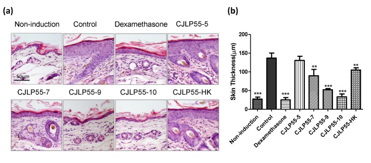
Fig. 2 Effect of orally administered CJLP55 on the thickness of the dorsal skin.(a) Histological analysis of the skin lesion after H&E staining. This is a representative image of five mice. (b) Epidermal thickness of the dorsal skin was measured. Data are shown as mean±SD of changes in the skin thickness of five mice (n=5). *p<0.05; **p<0.01; ***p<0.001 compared with control.
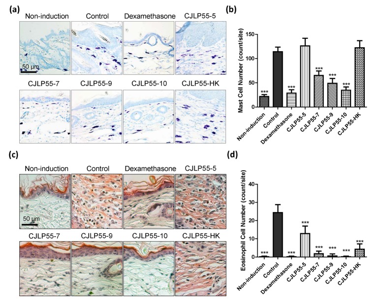
Fig. 3 Effects of orally administered CJLP55 on infiltration of mast cells and eosinophils in the inflammatory site.The paraffin block of the dorsal skin was sectioned and stained with toluidine blue (a) and congo red (c) for mast cells and eosinophils, respectively. The number of mast cells (b) and eosinophils (d) were counted under a microscope. Data are shown as mean±SD of five mice. *p<0.05; **p<0.01; ***p<0.001 compared with control.
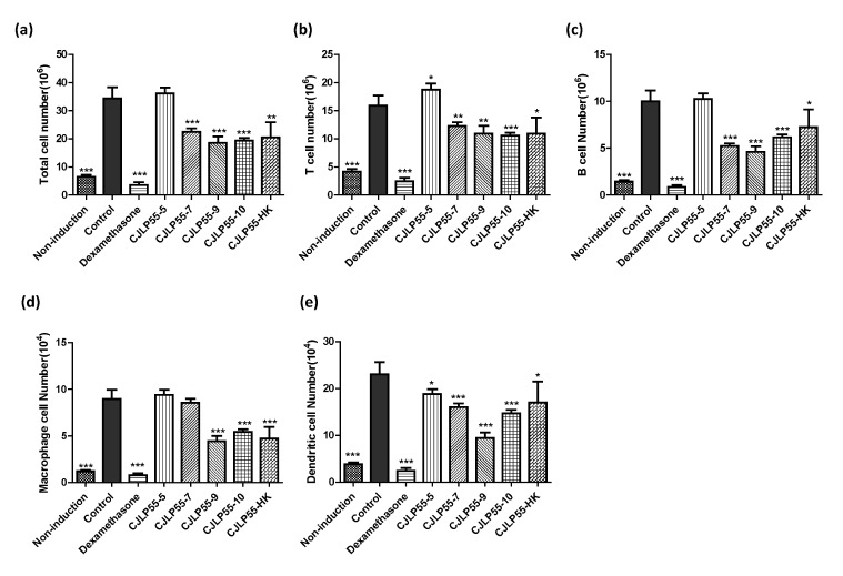
Fig. 4 Effects of orally administered CJLP55 on the axillary lymph nodes in NC/Nga mice.(a) Total cell number was measured from cell suspensions of the axillary lymph nodes. The number of T cells (b), B cells (c), macrophages (d), and dendritic cells (e) was analysed by flow cytometry. Data are shown as mean±SD of five mice. *p<0.05; **p<0.01; ***p<0.001 compared with control.
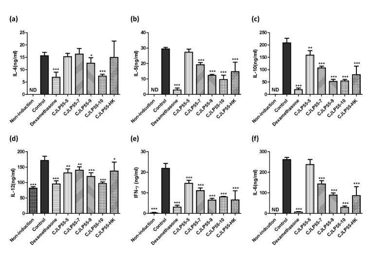
Fig. 5 Effects of orally administered CJLP55 on the cytokine production in the axillary lymph node cells.On day 56, axillary lymph node cells were isolated and cells were stimulated with 10 µgmL−1 of Dfb extract for 48 h. The concentration of IL-4 (a), IL-5 (b), IL-10 (c), IL-12 (d), IFN-γ (e), and IL-6 (f) was measured by ELISA. Data are shown as mean±SD of three independent experiments. *p<0.05; **p<0.01; ***p<0.001 compared with control.
Cited by 2 articles
-
Inhibitory effects of 2,6-di-
tert -butyl-4-hydroxymethylphenol on asthmatic responses to ovalbumin challenge in conscious guinea pigs
Seul-Yong Jeong, Ji-Yun Lee
Korean J Physiol Pharmacol. 2018;22(1):81-89. doi: 10.4196/kjpp.2018.22.1.81.Significance of Skin Barrier Dysfunction in Atopic Dermatitis
Byung Eui Kim, Donald Y.M. Leung
Allergy Asthma Immunol Res. 2018;10(3):207-215. doi: 10.4168/aair.2018.10.3.207.
Reference
-
2. Ogg G. Role of T cells in the pathogenesis of atopic dermatitis. Clin Exp Allergy. 2009; 39:310–316. PMID: 19040465.
Article3. Rodríguez JM, Murphy K, Stanton C, Ross RP, Kober OI, Juge N, Avershina E, Rudi K, Narbad A, Jenmalm MC, Marchesi JR, Collado MC. The composition of the gut microbiota throughout life, with an emphasis on early life. Microb Ecol Health Dis. 2015; 26:26050. PMID: 25651996.
Article4. West CE, Jenmalm MC, Prescott SL. The gut microbiota and its role in the development of allergic disease: a wider perspective. Clin Exp Allergy. 2015; 45:43–53. PMID: 24773202.
Article5. Hill C, Guarner F, Reid G, Gibson GR, Merenstein DJ, Pot B6, Morelli L, Canani RB, Flint HJ, Salminen S, Calder PC, Sanders ME. Expert consensus document. The International Scientific Association for Probiotics and Prebiotics consensus statement on the scope and appropriate use of the term probiotic. Nat Rev Gastroenterol Hepatol. 2014; 11:506–514. PMID: 24912386.6. Schiavi E, Barletta B, Butteroni C, Corinti S, Boirivant M, Di Felice G. Oral therapeutic administration of a probiotic mixture suppresses established Th2 responses and systemic anaphylaxis in a murine model of food allergy. Allergy. 2011; 66:499–508. PMID: 21058959.
Article7. Matsuzaki T, Takagi A, Ikemura H, Matsuguchi T, Yokokura T. Intestinal microflora: probiotics and autoimmunity. J Nutr. 2007; 137(3 Suppl 2):798S–802S. PMID: 17311978.
Article8. Won TJ, Kim B, Song DS, Lim YT, Oh ES, Lee DI, Park ES, Min H, Park SY, Hwang KW. Modulation of Th1/Th2 balance by Lactobacillus strains isolated from Kimchi via stimulation of macrophage cell line J774A.1 in vitro. J Food Sci. 2011; 76:H55–H61. PMID: 21535768.
Article9. Won TJ, Kim B, Lim YT, Song DS, Park SY, Park ES, Lee DI, Hwang KW. Oral administration of Lactobacillus strains from Kimchi inhibits atopic dermatitis in NC/Nga mice. J Appl Microbiol. 2011; 110:1195–1202. PMID: 21338447.
Article10. Yang G, Liu ZQ, Yang PC. Treatment of allergic rhinitis with probiotics: an alternative approach. N Am J Med Sci. 2013; 5:465–468. PMID: 24083221.
Article11. Matricardi PM, Bjorksten B, Bonini S, Bousquet J, Djukanovic R, Dreborg S, Gereda J, Malling HJ, Popov T, Raz E, Renz H, Wold A. EAACI Task Force 7. Microbial products in allergy prevention and therapy. Allergy. 2003; 58:461–471. PMID: 12757444.
Article12. Prescott SL, Björkstén B. Probiotics for the prevention or treatment of allergic diseases. J Allergy Clin Immunol. 2007; 120:255–262. PMID: 17544096.
Article13. Segawa S, Hayashi A, Nakakita Y, Kaneda H, Watari J, Yasui H. Oral administration of heat-killed Lactobacillus brevis SBC8803 ameliorates the development of dermatitis and inhibits immunoglobulin E production in atopic dermatitis model NC/Nga mice. Biol Pharm Bull. 2008; 31:884–889. PMID: 18451512.
Article14. Tobita K, Yanaka H, Otani H. Heat-treated Lactobacillus crispatus KT strains reduce allergic symptoms in mice. J Agric Food Chem. 2009; 57:5586–5590. PMID: 19469537.
Article15. Sunada Y, Nakamura S, Kamei C. Effect of Lactobacillus acidophilus strain L-55 on the development of atopic dermatitis-like skin lesions in NC/Nga mice. Int Immunopharmacol. 2008; 8:1761–1766. PMID: 18790088.
Article16. Matsuda H, Watanabe N, Geba GP, Sperl J, Tsudzuki M, Hiroi J, Matsumoto M, Ushio H, Saito S, Askenase PW, Ra C. Development of atopic dermatitis-like skin lesion with IgE hyperproduction in NC/Nga mice. Int Immunol. 1997; 9:461–466. PMID: 9088984.
Article17. Liu FT, Goodarzi H, Chen HY. IgE, mast cells, and eosinophils in atopic dermatitis. Clin Rev Allergy Immunol. 2011; 41:298–310. PMID: 21249468.
Article18. Lim HS, Ha H, Lee MY, Jin SE, Jeong SJ, Jeon WY, Shin NR, Sok DE, Shin HK. Saussurea lappa alleviates inflammatory chemokine production in HaCaT cells and house dust mite-induced atopic-like dermatitis in Nc/Nga mice. Food Chem Toxicol. 2014; 63:212–220. PMID: 24216625.
Article19. Matsuoka H, Maki N, Yoshida S, Arai M, Wang J, Oikawa Y, Ikeda T, Hirota N, Nakagawa H, Ishii A. A mouse model of the atopic eczema/dermatitis syndrome by repeated application of a crude extract of house-dust mite Dermatophagoides farinae. Allergy. 2003; 58:139–145. PMID: 12622745.
Article20. Yamamoto M, Haruna T, Yasui K, Takahashi H, Iduhara M, Takaki S, Deguchi M, Arimura A. A novel atopic dermatitis model induced by topical application with dermatophagoides farinae extract in NC/Nga mice. Allergol Int. 2007; 56:139–148. PMID: 17460441.
Article21. Imai Y, Yasuda K, Sakaguchi Y, Haneda T, Mizutani H, Yoshimoto T, Nakanishi K, Yamanishi K. Skin-specific expression of IL-33 activates group 2 innate lymphoid cells and elicits atopic dermatitis-like inflammation in mice. Proc Natl Acad Sci U S A. 2013; 110:13921–13926. PMID: 23918359.
Article22. Staumont-Sallé D, Fleury S, Lazzari A, Molendi-Coste O, Hornez N, Lavogiez C, Kanda A, Wartelle J, Fries A, Pennino D, Mionnet C, Prawitt J, Bouchaert E, Delaporte E, Glaichenhaus N, Staels B, Julia V, Dombrowicz D. CX3CL1 (fractalkine) and its receptor CX3CR1 regulate atopic dermatitis by controlling effector T cell retention in inflamed skin. J Exp Med. 2014; 211:1185–1196. PMID: 24821910.23. Kapp A. The role of eosinophils in the pathogenesis of atopic dermatitis--eosinophil granule proteins as markers of disease activity. Allergy. 1993; 48:1–5.24. Simon D, Braathen LR, Simon HU. Eosinophils and atopic dermatitis. Allergy. 2004; 59:561–570. PMID: 15147438.
Article25. Laouini D, Alenius H, Bryce P, Oettgen H, Tsitsikov E, Geha RS. IL-10 is critical for Th2 responses in a murine model of allergic dermatitis. J Clin Invest. 2003; 112:1058–1066. PMID: 14523043.
Article26. Fishman MA, Perelson AS. Th1/Th2 differentiation and cross-regulation. Bull Math Biol. 1999; 61:403–436. PMID: 17883225.
Article27. Grewe M, Bruijnzeel-Koomen CA, Schöpf E, Thepen T, Langeveld-Wildschut AG, Ruzicka T, Krutmann J. A role for Th1 and Th2 cells in the immunopathogenesis of atopic dermatitis. Immunol Today. 1998; 19:359–361. PMID: 9709503.
Article28. Toshitani A, Ansel JC, Chan SC, Li SH, Hanifin JM. Increased interleukin 6 production by T cells derived from patients with atopic dermatitis. J Invest Dermatol. 1993; 100:299–304. PMID: 8440909.
Article29. Gharagozlou M, Farhadi E, Khaledi M, Behniafard N, Sotoudeh S, Salari R, Darabi B, Fathi SM, Mahmoudi M, Aghamohammadi A, Amirzargar AA, Rezaei N. Association between the interleukin 6 genotype at position -174 and atopic dermatitis. J Investig Allergol Clin Immunol. 2013; 23:89–93.
- Full Text Links
-
- Actions
-
Cited
- CITED
-
- Close
- Share
-
- Similar articles
-
- Two Cases of Atopic Dermatitis Developing Ocular Complication and Immunological Disturbance
- Probiotics as an Immune Modulator for Allergic Disorders
- Intravenous immune globulin (i.v.IG) therapy in steroid-resistant atopic dermatitis
- A Study on the Cell - Mediated Immunity of Patients with Apopic Dermatitis
- Measurement of Atopic Dermatitis Disability
