The Thickness of Each Retinal Layer and Visual Acuity after Vitrectomy in Idiopathic Epiretinal Membrane
- Affiliations
-
- 1Department of Ophthalmology, Konyang University College of Medicine, Daejeon, Korea. astrix001@gmail.com
- 2Eyelove Clinic, Daejeon, Korea.
- 3Hwasun Country Public Health Center, Hwasun, Korea.
- KMID: 2376597
- DOI: http://doi.org/10.3341/jkos.2017.58.4.420
Abstract
- PURPOSE
In this study, we evaluated the thickness of each retinal layer using spectral-domain optical coherence tomography (OCT) and investigated the correlation between the thickness of each retinal layer and postoperative visual acuity in eyes with idiopathic epiretinal membrane (ERM).
METHODS
This retrospective study included 46 eyes from 46 patients with idiopathic ERM who underwent pars plana vitrectomy. Each retinal layer thickness was measured by spectral-domain OCT before operation and at 1, 3, and 6 months after operation. The thickness of each retinal layer was evaluated in the control group before the operation. We performed an analysis of the changes in thickness of each retinal layer at 6 months after operation and then investigated the correlation between the retinal layer thickness and visual improvement.
RESULTS
Preoperatively, the thickness of the retinal nerve fiber layer (RNFL) in the ERM group showed more increased compared with that in the control group, and the thickness of photoreceptors and retinal pigment epithelium were decreased compared to those in the control group. At 6 months after the operation, thickness changes were reduced at the RNFL, ganglion cell layer (GCL), inner plexiform layer (IPL), GCL-IPL complex, and outer plexiform layer, while the photoreceptor layer increased compared with the values preoperatively. Differences in the preoperative thickness of GCL between the two groups had a significant correlation with postoperative visual acuity (r = 0.477, p = 0.008).
CONCLUSIONS
Differences in preoperative thickness of the GCL between the two groups had a significant correlation with postoperative visual acuity. The greater was the thickness of the GCL, the worse was the visual outcome.
Keyword
MeSH Terms
Figure
-
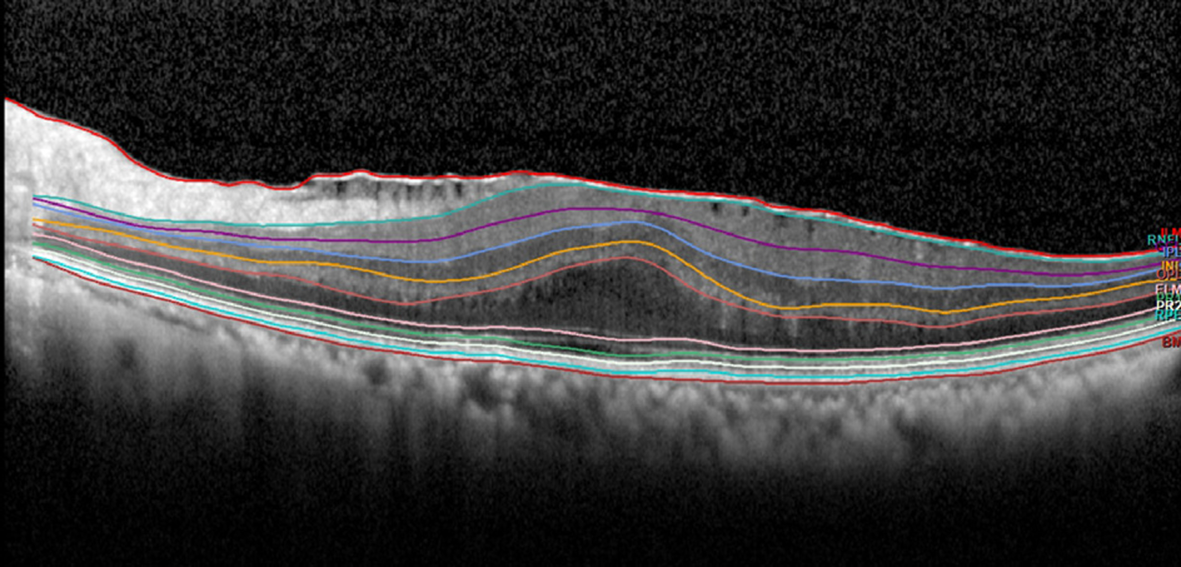
Figure 1. Representative image of retinal layer division determined by the new segmentation application of the spectralis optical coherence tomography in epiretinal membrane (Segmentation Technology; Heidelberg Engineering, Inc., Heidelberg, Germany). 10 layers: 1 = inner limiting membrane, 2 = retinal nerve fiber layer, 3 = ganglion cell layer, 4 = inner plexiform layer, 5 = inner nuclear layer, 6 = outer plexiform layer, 7 = external limiting membrane, 8 = photoreceptor, 9 = retinal pigment epithelium, 10 = Bruch's membrane.
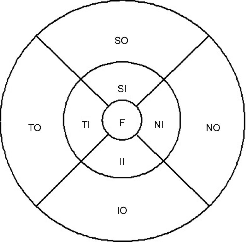
Figure 2. Early Treatment of Diabetic Retinopathy Study (ETDRS) subfield. Central circle: fovea (F). Inner ring: superior inner (SI) + nasal inner (NI) + inferior inner (II) + temporal inner (TI). Outer ring: superior outer (SO) + nasal outer (NO) + inferior outer (IO) + temporal outer (TO). ETDRS subfields within standard 1-, 3-, and 6-mm-diameter concentric circles at the right used for reporting retinal thickness.
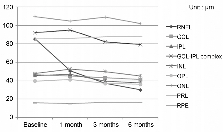
Figure 3. Longitudinal changes of the average foveal thick-ness of each retinal layer before and after epiretinal membrane surgery. RNFL =retinal nerve fiber layer; GCL =ganglion cell layer; IPL =inner plexiform layer; INL =inner nuclear layer; OPL =outer plexiform layer; ONL =outer nuclear lay-er; PRL = photoreceptor; RPE = retinal pigment epithelium.
Cited by 1 articles
-
Choroidal Thickness Changes Following Vitrectomy in Epiretinal Membrane Based on the Optical Coherence Tomography Pattern
Jun Min Park, Myeong In Yeom, Jung Min Park
J Korean Ophthalmol Soc. 2018;59(7):637-649. doi: 10.3341/jkos.2018.59.7.637.
Reference
-
References
1. Sidd RJ, Fine SL, Owens SL, Patz A. . Idiopathic preretinal gliosis. Am J Ophthalmol. 1982; 94:44–8.
Article2. Pearlstone AD. . The incidence of idiopathic preretinal macular gliosis. Ann Ophthalmol. 1985; 17:378–80.3. Mitchell P, Smith W, Chey T. . Prevalence and associations of epiretinal membranes. The Blue Mountains Eye Study, Australia. Ophthalmology. 1997; 104:1033–40.
Article4. Klein R, Klein BE, Wang Q, Moss SE. . The epidemiology of epiretinal membranes. Trans Am Ophthalmol Soc. 1994; 92:403–25. discussion 425-30.5. Machemer R. . The surgical removal of epiretinal macular membranes (macular puckers) (author's transl). Klin Monbl Augenheilkd. 1978; 173:36–42.6. McDonald HR, Verre WP, Aaberg TM. . Surgical management of idiopathic epiretinal membranes. Ophthalmology. 1986; 93:978–83.
Article7. de Bustros S, Thompson JT, Michels RG. . Nuclear sclerosis after vitrectomy for idiopathic epiretinal membranes. Am J Ophthalmol. 1988; 105:160–4.8. Margherio RR, Cox MS Jr, Trese MT. . Removal of epimacular membranes. Ophthalmology. 1985; 92:1075–83.
Article9. Mitamura Y, Hirano K, Baba T, Yamamoto S. . Correlation of visual recovery with presence of photoreceptor inner/outer segment junction in optical coherence images after epiretinal membrane surgery. Br J Ophthalmol. 2009; 93:171–5.
Article10. Inoue M, Morita S, Watanabe Y. . Preoperative inner seg-ment/outer segment junction in spectral-domain optical coherence tomography as a prognostic factor in epiretinal membrane surgery. Retina. 2011; 31:1366–72.
Article11. Shimozono M, Oishi A, Hata M. . The significance of cone outer segment tips as a prognostic factor in epiretinal membrane surgery. Am J Ophthalmol. 2012; 153:698–704. 704.e1.
Article12. Lee EK, Yu HG. . Ganglion cell-inner plexiform layer thickness af-ter epiretinal membrane surgery: a spectral-domain optical coher-ence tomography study. Ophthalmology. 2014; 121:1579–87.13. Okamoto F, Sugiura Y, Okamoto F. . Inner nuclear layer thick-ness as a prognostic factor for metamorphopsia after epiretinal membrane surgery. Retina. 2015; 35:2107–14.
Article14. Lee JY, Kim HC. . Ganglion cell layer thickness after anti-vascular endothelial growth factor treatment in retinal vein occlusion. J Korean Ophthalmol Soc. 2016; 57:63–70.
Article15. Arichika S, Hangai M, Yoshimura N. . Correlation between thickening of the inner and outer retina and visual acuity in patients with epiretinal membrane. Retina. 2010; 30:503–8.
Article16. Koo HC, Rhim WI, Lee EK. . Morphologic and functional association of retinal layers beneath the epiretinal membrane with spec-tral-domain optical coherence tomography in eyes without photo-receptor abnormality. Graefes Arch clin Exp Ophthalmol. 2012; 250:491–8.
Article17. Rice TA, De Bustros S, Michels RG. . Prognostic factors in vi-trectomy for epiretinal membranes of the macula. Ophthalmology. 1986; 93:602–10.
Article18. de Bustros S, Rice TA, Michels RG. . Vitrectomy for macular pucker. Use after treatment of retinal tears or retinal detachment. Arch Ophthalmol. 1988; 106:758–60.19. Kwon SI, Ko SJ, Park IW. . The clinical course of the idiopathic epi-retinal membrane after surgery. Korean J Ophthalmol. 2009; 23:249–52.
Article20. Kim J, Rhee KM, Woo SJ. . Long-term temporal changes of macular thickness and visual outcome after vitrectomy for idio-pathic epiretinal membrane. Am J Ophthalmol. 2010; 150:701–9. e1.
Article21. Hwang DJ, Na KI, Kwon SI, Park IW. . Long-term changes in visual acuity and foveal thickness after vitrectomy for idiopathic epi-retinal membrane. J Korean Ophthalmol Soc. 2012; 53:434–9.
Article22. Wolf-Schnurrbusch UE, Ceklic L, Brinkmann CK. . Macular thickness measurements in healthy eyes using six different optical coherence tomography instruments. Invest Ophthalmol Vis Sci. 2009; 50:3432–7.
Article23. Ctori I, Huntjens B. . Repeatability of foveal measurements using spectralis optical coherence tomography segmentation software. PLoS ONE. 2015; 10:e0129005.
Article
- Full Text Links
-
- Actions
-
Cited
- CITED
-
- Close
- Share
-
- Similar articles
-
- Evaluation of Each Retinal Layer Thickness According to Preoperative OCT Patterns after Idiopathic ERM Removal
- Visual and Structural Differences in Idiopathic Epiretinal Membrane According to the Presence of Retinoschisis
- Long-Term Changes in Visual Acuity and Foveal Thickness after Vitrectomy for Idiopathic Epiretinal Membrane
- The Clinical Course of the Idiopathic Epiretinal Membrane After Surgery
- Comparison of Visual Acuity and Retinal Thickness According to Membranectomy in Idiopathic Epiretinal Membrane
