Temporal Evolution of a Chronic Expanding Organizing Hematoma on MRI, Including Functional MR Imaging Techniques: a Case Report
- Affiliations
-
- 1Department of Radiology, Samsung Medical Center, Sungkyunkwan University School of Medicine, Seoul, Korea. youngcheol.yoon@samsung.com
- 2Department of Pathology and Translational Genomics, Samsung Medical Center, Sungkyunkwan University School of Medicine, Seoul, Korea.
- KMID: 2376269
- DOI: http://doi.org/10.13104/imri.2017.21.1.43
Abstract
- Chronic expanding organizing hematoma (CEH) occasionally mimics a soft tissue tumor on MRI, which becomes more problematic in patients with a history of surgical resection for musculoskeletal malignancy. Herein, we present a case of CEH which we were able to differentiate from recurrent tumor through MRI follow-up, including diffusion-weighted imaging (DWI) and dynamic contrast enhanced (DCE) imaging. A 66-year-old male visited our institution under suspicion of recurrent leiomyosarcoma of the thigh, 19 months after surgery and radiation therapy. Due to inconclusive results, three US-guided biopsies and 6 MRI examinations were performed over 2 years. In the end, we could diagnose a CEH using conventional and functional MRI techniques, and it was histopathologically confirmed after surgical resection. A CEH may occur remotely after an initiating event, and it may persist and expand over several years. Functional MR sequences, in addition to conventional sequences, are helpful in differentiating CEH from malignant neoplasms.
Keyword
MeSH Terms
Figure
-
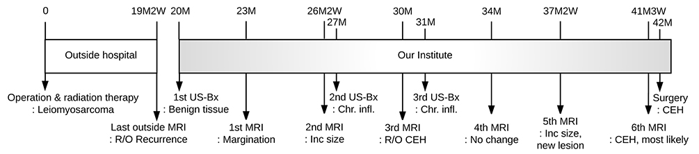
Fig. 1 Overview of the patient's clinical course. CEH = chronic expanding organizing hematoma; Chr. infl. = chronic inflammation; Inc size = increased size; M = month; US-Bx = US-guided biopsy; W = week
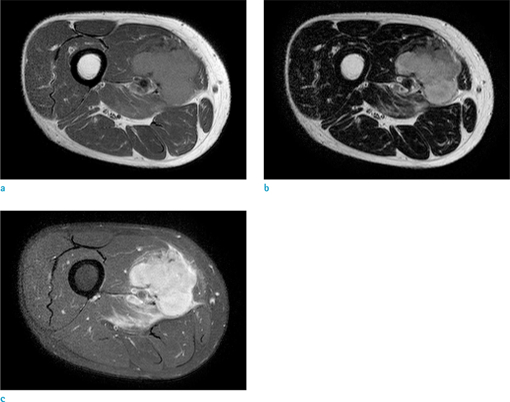
Fig. 2 Initial MRI obtained at the previous hospital. T1-weighted image (a), T2-weighted image (b), and T1-weighted contrast-enhanced image (c). There is a well-circumscribed mass with lobulated contours in the mid-thigh. The pathologic diagnosis was leiomyosarcoma.
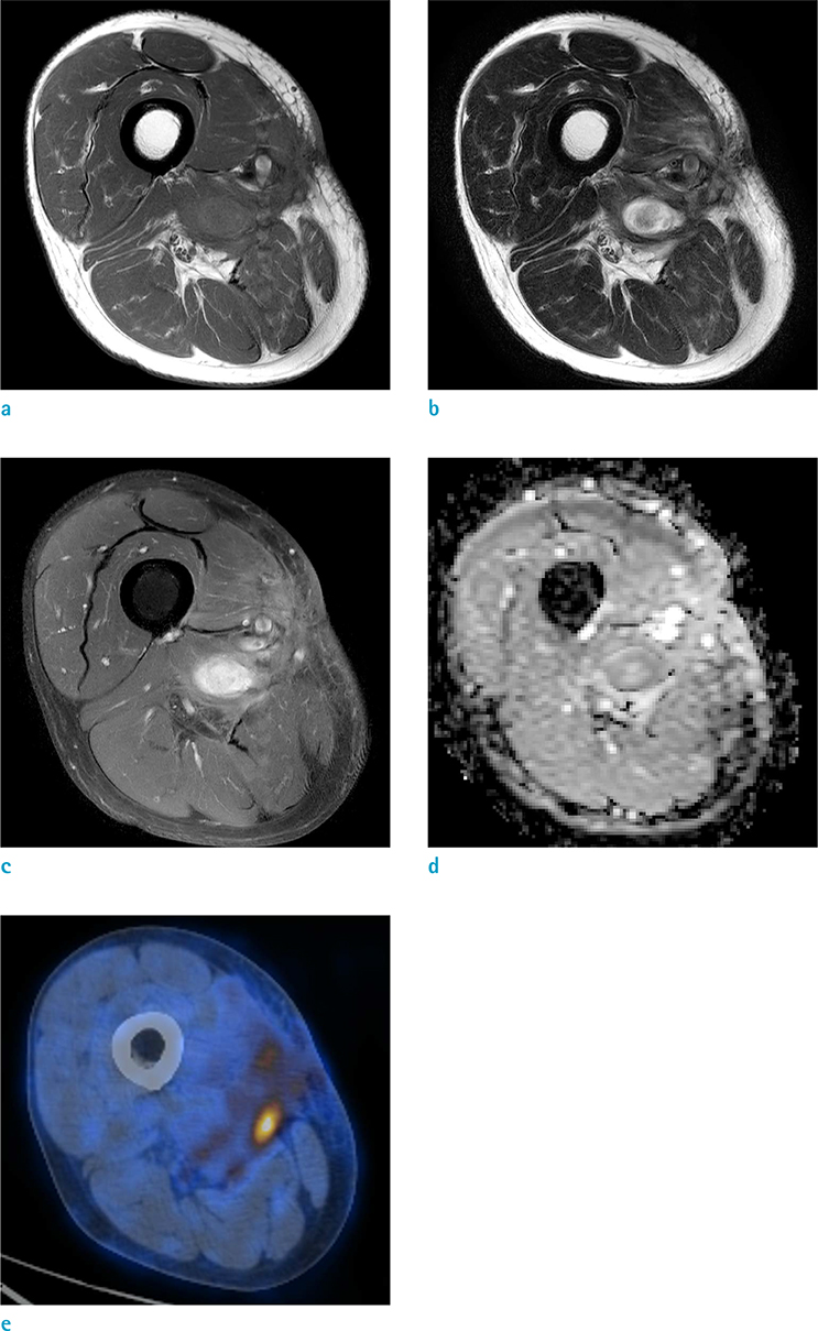
Fig. 3 Second MRI obtained at our institute. T1-weighted image (a), T2-weighted image (b), T1-weighted contrast-enhanced image (c), apparent diffusion coefficient (ADC) map (d), and PET-CT obtained two weeks before (e). An enhancing nodular lesion in the operative bed is seen. This lesion shows an increase in size, compared to the previous MRI examination (not shown). The ADC map does not show diffusion restriction.
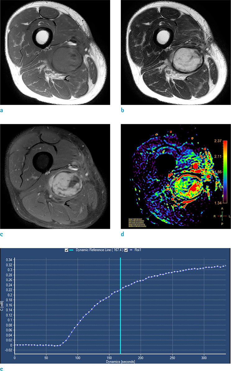
Fig. 4 Sixth (last) MRI obtained at our institute. T1-weighted image (a), T2-weighted image (b), T1-weighted contrast-enhanced image (c), apparent diffusion coefficient (ADC) map (d), and dynamic enhancement curve (e). The growing mass now shows a distinct hypointense rim on T2-weighted images, which is a characteristic finding of chronic expanding organizing hematoma. The high ADC value and non-arterial enhancement curve favor benignity.
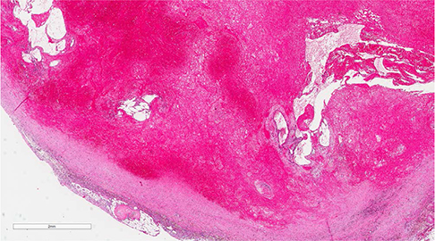
Fig. 5 Low-power microscopic photograph of the surgical specimen. There is chronic inflammation and granulation tissue, with a surrounding thick fibrous capsule. Fibrinous exudates with hemorrhage, dilated vessels, and neovascularization were seen within the lesion. Pathology report confirmed the diagnosis of chronic expanding organizing hematoma.
Reference
-
1. Reid JD, Kommareddi S, Lankerani M, Park MC. Chronic expanding hematomas. A clinicopathologic entity. JAMA. 1980; 244:2441–2442.2. Sreenivas M, Nihal A, Ettles DF. Chronic haematoma or soft-tissue neoplasm? A diagnostic dilemma. Arch Orthop Trauma Surg. 2004; 124:495–497.3. Oka K, Yakushiji T, Sato H, et al. Ability of diffusion-weighted imaging for the differential diagnosis between chronic expanding hematomas and malignant soft tissue tumors. J Magn Reson Imaging. 2008; 28:1195–1200.4. Aoki T, Nakata H, Watanabe H, et al. The radiological findings in chronic expanding hematoma. Skeletal Radiol. 1999; 28:396–401.5. Park JC, Ahn JS, Kwon DH, Kwun BD. Growing organized hematomas following gamma knife radiosurgery for cerebral arteriovenous malformation: five cases of surgical excision. J Korean Neurosurg Soc. 2015; 58:83–88.6. Nishida Y, Kobayashi E, Kubota D, et al. Chronic expanding hematoma with a significantly high fluorodeoxyglucose uptake on 18F-fluorodeoxyglucose positron emission tomography, mimicking a malignant soft tissue tumor: a case report. J Med Case Rep. 2014; 8:349.7. Kasraeian S, Allison DC, Ahlmann ER, Fedenko AN, Menendez LR. A comparison of fine-needle aspiration, core biopsy, and surgical biopsy in the diagnosis of extremity soft tissue masses. Clin Orthop Relat Res. 2010; 468:2992–3002.8. Kim JI, Lee IS, Song YS, Park SK, Choi KU, Song JW. Short-term follow-up MRI after unplanned resection of malignant soft-tissue tumours; quantitative measurements on dynamic contrast enhanced and diffusion-weighted MR images. Br J Radiol. 2016; 89:20160302.9. Del Grande F, Subhawong T, Weber K, Aro M, Mugera C, Fayad LM. Detection of soft-tissue sarcoma recurrence: added value of functional MR imaging techniques at 3.0 T. Radiology. 2014; 271:499–511.10. Surov A, Nagata S, Razek AA, Tirumani SH, Wienke A, Kahn T. Comparison of ADC values in different malignancies of the skeletal musculature: a multicentric analysis. Skeletal Radiol. 2015; 44:995–1000.
- Full Text Links
-
- Actions
-
Cited
- CITED
-
- Close
- Share
-
- Similar articles
-
- A Case of Orbital Organizing Hematoma Presenting as a Chalazion
- A Case of Organizing Hematoma of the Nasal Septum
- Unilateral Chronic Organizing Hematoma after Breast Explantation Mimicking Chest Wall Tumor: a Case Report with Imaging Features
- Three Cases of Organizing Hematoma in the Maxillary Sinus
- A Case of Surgical Management for Orbital Organizing Hematoma from Orbital Varix
