Korean J Ophthalmol.
2016 Dec;30(6):451-458. 10.3341/kjo.2016.30.6.451.
Macular Ganglion Cell Layer Assessment to Detect Glaucomatous Central Visual Field Progression
- Affiliations
-
- 1Department of Ophthalmology, Asan Medical Center, University of Ulsan College of Medicine, Seoul, Korea. sungeye@gmail.com
- KMID: 2374003
- DOI: http://doi.org/10.3341/kjo.2016.30.6.451
Abstract
- PURPOSE
To investigate the use of ganglion cell inner plexiform layer (GC-IPL) thickness, as measured by spectral domain optical coherence tomography, to detect central visual field (VF) progression.
METHODS
This study included 384 eyes from 384 patients (219 preperimetric and 165 perimetric glaucomatous eyes; average follow-up, 4.3 years). Photographic assessment of retinal nerve fiber layer (RNFL) and serial VF analysis were performed to detect glaucoma progression in the central (within 10°) area. Study inclusion required at least five serial spectral domain optical coherence tomography exams at different visits. The long-term test-retest variability of average GC-IPL thicknesses was calculated in 110 stable preperimetric glaucomatous eyes. The sensitivity and specificity of GC-IPL measurements for the detection of central VF progression were calculated in an event-based analysis using the calculated variability as a cut-off and were compared with those of central RNFL photographic assessment.
RESULTS
The intersession test-retest variability, defined as the 95% confidence interval, was 1.76 µm for average GC-IPL thickness. The sensitivity and specificity of the average GC-IPL thickness for detecting central VF progression were 60.7% and 78.9%, respectively. Among six sectors, the inferonasal GC-IPL sector showed the highest sensitivity (53.6%). The sensitivity of the ≥1 sector GC-IPL to detect central VF progression was significantly higher than that of central RNFL photographic progression (p = 0.013). Other GC-IPL parameters showed comparable sensitivity and specificity to detect central VF progression compared with RNFL photographic progression.
CONCLUSIONS
Serial GC-IPL measurements show comparable performance in the detection of central glaucomatous VF progression to RNFL photographic assessment.
MeSH Terms
Figure
-
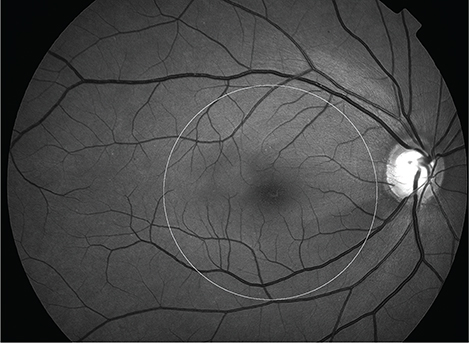
Fig. 1 The central circle (6-mm diameter) corresponds to the ganglion cell inner plexiform layer map size overlapping the redfree retinal nerve fiber layer photograph to determine central retinal nerve fiber layer progression.
Reference
-
1. Burgansky-Eliash Z, Wollstein G, Chu T, et al. Optical coherence tomography machine learning classifiers for glaucoma detection: a preliminary study. Invest Ophthalmol Vis Sci. 2005; 46:4147–4152.2. Lalezary M, Medeiros FA, Weinreb RN, et al. Baseline optical coherence tomography predicts the development of glaucomatous change in glaucoma suspects. Am J Ophthalmol. 2006; 142:576–582.3. Leung CK, Chan WM, Yung WH, et al. Comparison of macular and peripapillary measurements for the detection of glaucoma: an optical coherence tomography study. Ophthalmology. 2005; 112:391–400.4. Manassakorn A, Nouri-Mahdavi K, Caprioli J. Comparison of retinal nerve fiber layer thickness and optic disk algorithms with optical coherence tomography to detect glaucoma. Am J Ophthalmol. 2006; 141:105–115.5. Na JH, Sung KR, Baek SH, et al. Rates and patterns of macular and circumpapillary retinal nerve fiber layer thinning in preperimetric and perimetric glaucomatous eyes. J Glaucoma. 2015; 24:278–285.6. Na JH, Sung KR, Lee JR, et al. Detection of glaucomatous progression by spectral-domain optical coherence tomography. Ophthalmology. 2013; 120:1388–1395.7. Na JH, Sung KR, Baek S, et al. Detection of glaucoma progression by assessment of segmented macular thickness data obtained using spectral domain optical coherence tomography. Invest Ophthalmol Vis Sci. 2012; 53:3817–3826.8. Seong M, Sung KR, Choi EH, et al. Macular and peripapillary retinal nerve fiber layer measurements by spectral domain optical coherence tomography in normal-tension glaucoma. Invest Ophthalmol Vis Sci. 2010; 51:1446–1452.9. Sung KR, Wollstein G, Kim NR, et al. Macular assessment using optical coherence tomography for glaucoma diagnosis. Br J Ophthalmol. 2012; 96:1452–1455.10. Shin HY, Park HY, Jung KI, et al. Glaucoma diagnostic ability of ganglion cell-inner plexiform layer thickness differs according to the location of visual field loss. Ophthalmology. 2014; 121:93–99.11. Leung CK, Ye C, Weinreb RN, et al. Impact of age-related change of retinal nerve fiber layer and macular thicknesses on evaluation of glaucoma progression. Ophthalmology. 2013; 120:2485–2492.12. Sung KR, Sun JH, Na JH, et al. Progression detection capability of macular thickness in advanced glaucomatous eyes. Ophthalmology. 2012; 119:308–313.13. Sung KR, Kim DY, Park SB, Kook MS. Comparison of retinal nerve fiber layer thickness measured by Cirrus HD and Stratus optical coherence tomography. Ophthalmology. 2009; 116:1264–1270.e1.14. Heijl A, Leske MC, Bengtsson B, et al. Measuring visual field progression in the Early Manifest Glaucoma Trial. Acta Ophthalmol Scand. 2003; 81:286–293.15. Sung KR, Cho JW, Lee S, et al. Characteristics of visual field progression in medically treated normal-tension glaucoma patients with unstable ocular perfusion pressure. Invest Ophthalmol Vis Sci. 2011; 52:737–743.16. Omodaka K, Takada N, Takahashi H, Nakazawa T. Regional structural vulnerability of the macula in patients with normal tension glaucoma. Clin Experiment Ophthalmol. 2015; 43:89–90.17. Lee JR, Sung KR, Na JH, et al. Discrepancy between optic disc and nerve fiber layer assessment and optical coherence tomography in detecting glaucomatous progression. Jpn J Ophthalmol. 2013; 57:546–552.18. Na JH, Sung KR, Baek S, et al. Progression of retinal nerve fiber layer thinning in glaucoma assessed by Cirrus optical coherence tomography-guided progression analysis. Curr Eye Res. 2013; 38:386–395.19. Moon BG, Sung KR, Cho JW, et al. Glaucoma progression detection by retinal nerve fiber layer measurement using scanning laser polarimetry: event and trend analysis. Korean J Ophthalmol. 2012; 26:174–181.20. Carpineto P, Aharrh-Gnama A, Ciciarelli V, et al. Reproducibility and repeatability of ganglion cell-inner plexiform layer thickness measurements in healthy subjects. Ophthalmologica. 2014; 232:163–169.21. Kim KE, Yoo BW, Jeoung JW, Park KH. Long-term reproducibility of macular ganglion cell analysis in clinically stable glaucoma patients. Invest Ophthalmol Vis Sci. 2015; 56:4857–4864.22. Takayama K, Hangai M, Durbin M, et al. A novel method to detect local ganglion cell loss in early glaucoma using spectral-domain optical coherence tomography. Invest Ophthalmol Vis Sci. 2012; 53:6904–6913.
- Full Text Links
-
- Actions
-
Cited
- CITED
-
- Close
- Share
-
- Similar articles
-
- RNFL and Ganglion Cell Complex Thickness in Normal Hemifield According to the Severity of Glaucoma
- Longitudinal Analysis of Retinal Nerve Fiber Layer Thickness With GDx-VCC in Glaucoma Suspect
- Hierarchical Cluster Analysis of Peripapillary Retinal Nerve Fiber Layer Damage and Macular Ganglion Cell Loss in Open Angle Glaucoma
- Correlation Between Nocturnal Dip and Progression of Glaucoma
- Visual Field Changes after Internal Limiting Membrane Peeling in Glaucoma Patients with Epiretinal Membrane
