Clinical and Anterior Segment Anatomical Features in Primary Angle Closure Subgroups Based on Configurations of Iris Root Insertion
- Affiliations
-
- 1Department of Ophthalmology, University of Ulsan College of Medicine, Seoul, Korea. sungeye@gmail.com
- 2Department of Clinical Epidemiology and Biostatistics, Asan Medical Center, University of Ulsan College of Medicine, Seoul, Korea.
- KMID: 2373976
- DOI: http://doi.org/10.3341/kjo.2016.30.3.206
Abstract
- PURPOSE
To compare the clinical and anterior segment anatomical features in primary angle closure sub-groups based on configurations of iris root insertion.
METHODS
Primary angle closure patients were imaged using anterior segment optical coherence tomography. Anterior chamber depth, iris curvature, iris thickness (IT) at the scleral spur and 500, 750, and 1,500 µm from the scleral spur (IT(0), IT(500), IT(750), and IT(1500)), lens vault, iris area, angle opening distance (AOD(500)), angle recess area (ARA(750)), and trabecular iris space area (TISA(750)) were measured. Iris root insertion was categorized into a non-basal insertion group (NBG) and basal insertion group (BG).
RESULTS
In total, 43 eyes of 39 participants belonged to the NBG and 89 eyes of 53 participants to the BG. The mean age of participants was greater in the NBG than the BG (62.7 ± 5.7 vs. 59.8 ± 7.3 years, p = 0.043), and the baseline intraocular pressure was higher in the BG than the NBG (16.4 ± 4.4 vs. 14.9 ± 3.3 mmHg, p = 0.037). The BG showed a greater IT(0) (0.265 ± 0.04 vs. 0.214 ± 0.03 mm, p < 0.001) and iris area (1.59 ± 0.24 vs. 1.52 ± 0.27 mm2, p = 0.045), lower ARA(750) (0.112 ± 0.08 vs. 0.154 ± 0.08 mm2, p = 0.017) and AOD(500) (0.165 ± 0.07 vs. 0.202 ± 0.08 mm, p = 0.014) compared to the NBG.
CONCLUSIONS
The BG had a narrower anterior chamber angle, thicker peripheral iris, and higher pretreatment intraocular pressure.
Keyword
MeSH Terms
-
Anterior Eye Segment/*diagnostic imaging
Female
Glaucoma, Angle-Closure/diagnosis/physiopathology/*surgery
Gonioscopy
Humans
*Intraocular Pressure
Iridectomy/*methods
Iris/diagnostic imaging/*surgery
Lens, Crystalline/diagnostic imaging
Male
Middle Aged
Prospective Studies
Tomography, Optical Coherence/*methods
Figure
-

Fig. 1 Anterior segment parameters measured by anterior segment optical coherence tomography and calculated using the Image J software ver. 1.46 (National Institutes of Health, Bethesda, MD, USA). AOD500, 750 = angle opening distance; SS = scleral spur; IT0, 500, 750, 1500 = iris thickness; AA = anterior chamber area; ACD = anterior chamber depth; LV = lens vault; PD = pupil distance; IA = iris area; IC = iris curvature; TISA750 = trabecular iris space area; ARA750 = angle recess area.
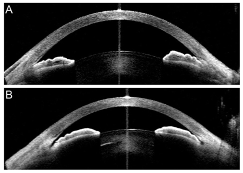
Fig. 2 Location of iris root insertion. (A) Basal insertion and (B) non-basal insertion.
Cited by 1 articles
-
Effects of Choroidal Thickness on Refractive Outcome Following Cataract Surgery in Primary Angle Closure
Woo Keun Song, Kyung Rim Sung, Joong Won Shin, Junki Kwon
Korean J Ophthalmol. 2018;32(5):382-390. doi: 10.3341/kjo.2017.0129.
Reference
-
1. Congdon N, Wang F, Tielsch JM. Issues in the epidemiology and population-based screening of primary angle-closure glaucoma. Surv Ophthalmol. 1992; 36:411–423.2. Foster PJ, Baasanhu J, Alsbirk PH, et al. Glaucoma in Mongolia: a population-based survey in Hovsgol province, northern Mongolia. Arch Ophthalmol. 1996; 114:1235–1241.3. Foster PJ, Johnson GJ. Glaucoma in China: how big is the problem? Br J Ophthalmol. 2001; 85:1277–1282.4. Lowe RF. Aetiology of the anatomical basis for primary angle-closure glaucoma: biometrical comparisons between normal eyes and eyes with primary angle-closure glaucoma. Br J Ophthalmol. 1970; 54:161–169.5. Sihota R, Lakshmaiah NC, Agarwal HC, et al. Ocular parameters in the subgroups of angle closure glaucoma. Clin Experiment Ophthalmol. 2000; 28:253–258.6. George R, Paul PG, Baskaran M, et al. Ocular biometry in occludable angles and angle closure glaucoma: a population based survey. Br J Ophthalmol. 2003; 87:399–402.7. Aung T, Nolan WP, Machin D, et al. Anterior chamber depth and the risk of primary angle closure in 2 East Asian populations. Arch Ophthalmol. 2005; 123:527–532.8. Cheon MH, Sung KR, Choi EH, et al. Effect of age on anterior chamber angle configuration in Asians determined by anterior segment optical coherence tomography: clinic-based study. Acta Ophthalmol. 2010; 88:e205–e210.9. Kim DY, Sung KR, Kang SY, et al. Characteristics and reproducibility of anterior chamber angle assessment by anterior-segment optical coherence tomography. Acta Ophthalmol. 2011; 89:435–441.10. Wang BS, Narayanaswamy A, Amerasinghe N, et al. Increased iris thickness and association with primary angle closure glaucoma. Br J Ophthalmol. 2011; 95:46–50.11. Wang B, Sakata LM, Friedman DS, et al. Quantitative iris parameters and association with narrow angles. Ophthalmology. 2010; 117:11–17.12. Sung KR, Lee KS, Hong JW. Baseline anterior segment parameters associated with the long-term outcome of laser peripheral iridotomy. Curr Eye Res. 2015; 40:1128–1133.13. Lee RY, Kasuga T, Cui QN, et al. Association between baseline iris thickness and prophylactic laser peripheral iridotomy outcomes in primary angle-closure suspects. Ophthalmology. 2014; 121:1194–1202.14. Foster PJ, Buhrmann R, Quigley HA, Johnson GJ. The definition and classification of glaucoma in prevalence surveys. Br J Ophthalmol. 2002; 86:238–242.15. Lee KY, Rensch F, Aung T, et al. Peripapillary atrophy after acute primary angle closure. Br J Ophthalmol. 2007; 91:1059–1061.16. Lee Y, Sung KR, Na JH, Sun JH. Dynamic changes in anterior segment (AS) parameters in eyes with primary angle closure (PAC) and PAC glaucoma and open-angle eyes assessed using AS optical coherence tomography. Invest Ophthalmol Vis Sci. 2012; 53:693–697.17. Sakata LM, Lavanya R, Friedman DS, et al. Assessment of the scleral spur in anterior segment optical coherence tomography images. Arch Ophthalmol. 2008; 126:181–185.18. Nongpiur ME, He M, Amerasinghe N, et al. Lens vault, thickness, and position in Chinese subjects with angle closure. Ophthalmology. 2011; 118:474–479.19. Baek S, Sung KR, Sun JH, et al. A hierarchical cluster analysis of primary angle closure classification using anterior segment optical coherence tomography parameters. Invest Ophthalmol Vis Sci. 2013; 54:848–853.20. Lee KS, Sung KR, Shon K, et al. Longitudinal changes in anterior segment parameters after laser peripheral iridotomy assessed by anterior segment optical coherence tomography. Invest Ophthalmol Vis Sci. 2013; 54:3166–3170.21. Sun JH, Sung KR, Yun SC, et al. Factors associated with anterior chamber narrowing with age: an optical coherence tomography study. Invest Ophthalmol Vis Sci. 2012; 53:2607–2610.22. Han S, Sung KR, Lee KS, Hong JW. Outcomes of laser peripheral iridotomy in angle closure subgroups according to anterior segment optical coherence tomography parameters. Invest Ophthalmol Vis Sci. 2014; 55:6795–6801.23. Wang YE, Li Y, Wang D, et al. Comparison of iris insertion classification among American caucasian and ethnic Chinese using ultrasound biomicroscopy. Invest Ophthalmol Vis Sci. 2013; 54:3837–3843.24. Ku JY, Nongpiur ME, Park J, et al. Qualitative evaluation of the iris and ciliary body by ultrasound biomicroscopy in subjects with angle closure. J Glaucoma. 2014; 23:583–588.25. Jiang Y, He M, Huang W, et al. Qualitative assessment of ultrasound biomicroscopic images using standard photographs: the liwan eye study. Invest Ophthalmol Vis Sci. 2010; 51:2035–2042.26. Chen HJ, Wang X, Yan YJ, Wu LL. Postiridotomy ultrasound biomicroscopy features in the fellow eye of Chinese patients with acute primary angle-closure and chronic primary angle-closure glaucoma. J Glaucoma. 2015; 24:233–237.27. Kumar RS, Tantisevi V, Wong MH, et al. Plateau iris in Asian subjects with primary angle closure glaucoma. Arch Ophthalmol. 2009; 127:1269–1272.28. Aptel F, Denis P. Optical coherence tomography quantitative analysis of iris volume changes after pharmacologic mydriasis. Ophthalmology. 2010; 117:3–10.29. Quigley HA. The iris is a sponge: a cause of angle closure. Ophthalmology. 2010; 117:1–2.
- Full Text Links
-
- Actions
-
Cited
- CITED
-
- Close
- Share
-
- Similar articles
-
- Measurement of The Depth and Angle of The Peripheral Anterior Chamber and Iris Thickness by Ultrasonographic Biomicroscopy
- Ultrasound Biomicroscopic Assessment of the Changes in Angle Relationships after Laser Iridotomy in Primary Angle-Closure Glaucoma
- Clinical Experiences of Iridocycloretraction Operation in Chronic Angle Closure Glaucoma
- Ultrasound biomicroscopic dimensions of the anterior chamber in angle-closure glaucoma patients
- Ultrasound Biomicroscopic Changes after Laser Iridotomy or Trabeculectomy in Angle-closure Glaucoma
