J Korean Med Sci.
2016 Jun;31(6):1007-1008. 10.3346/jkms.2016.31.6.1007.
Subcutaneous Gas of the Perineal Area: Radiographic Signs of Fournier's Gangrene
- Affiliations
-
- 1Department of Trauma, Chi Mei Medical Center, Tainan, Taiwan.
- 2Department of Physical Therapy, Shu Zen College of Medicine and Management, Tainan, Taiwan.
- 3Division of Geriatrics and Gerontology, Department of Internal Medicine, Kaohsiung Medical University Hospital, Kaohsiung Medical University, Kaohsiung, Taiwan.
- 4Department of Neurology, Kaohsiung Medical University Hospital, Kaohsiung Medical University, Kaohsiung, Taiwan.
- 5Department of Intensive Care Medicine, Chi Mei Medical Center, Liouying, Tainan, Taiwan. ccm870958@yahoo.com.tw
- 6Department of Nursing, Min-Hwei College of Health Care Management, Tainan, Taiwan.
- KMID: 2373714
- DOI: http://doi.org/10.3346/jkms.2016.31.6.1007
Abstract
- No abstract available.
Figure
-
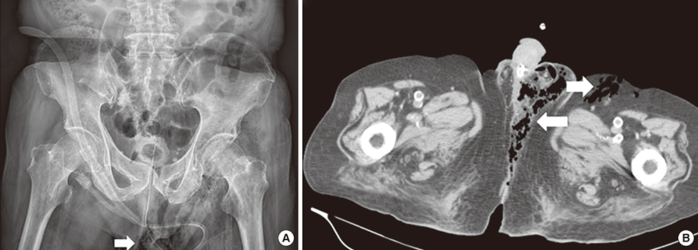
Fig. 1 Radiography images of the pelvis. (A) Subcutaneous emphysema over left perineal region (arrow). (B) Computed tomography of abdomen showing the presence of air over left perinea, inguinal and lower abdominal wall region (arrows).
Reference
-
1. Sorensen MD, Krieger JN, Rivara FP, Broghammer JA, Klein MB, Mack CD, Wessells H. Fournier's Gangrene: population based epidemiology and outcomes. J Urol. 2009; 181:2120–2126.2. Yilmazlar T, Ozturk E, Ozguc H, Ercan I, Vuruskan H, Oktay B. Fournier's gangrene: an analysis of 80 patients and a novel scoring system. Tech Coloproctol. 2010; 14:217–223.3. Levenson RB, Singh AK, Novelline RA. Fournier gangrene: role of imaging. Radiographics. 2008; 28:519–528.
- Full Text Links
-
- Actions
-
Cited
- CITED
-
- Close
- Share
-
- Similar articles
-
- Two cases of Fournier's gangrene
- Fournier's Gangrene after Excision of a Thrombosed Hemorrhoid
- Fournier's Gangrene: A report of one case
- Fournier’s Gangrene in a Female Infant
- Medial Femoral Circumflex Artery Perforator Based Fasciocutaneous Flap Aided in Healing of Scrotal Defect due to Fournier Gangrene
