Acute Myeloid Leukemia With MLL Rearrangement and CD4+/CD56+ Expression can be Misdiagnosed as Blastic Plasmacytoid Dendritic Cell Neoplasm: Two Case Reports
- Affiliations
-
- 1Department of Laboratory Medicine, Pusan National University Yangsan Hospital, Yangsan, Korea. iskim0710@gmail.com
- 2Research Institute for Convergence of Biomedical Science and Technology, Pusan National University Yangsan Hospital, Yangsan, Korea.
- 3Department of Laboratory Medicine, Haeundae Paik Hospital, Inje University College of Medicine, Busan, Korea. jeong418@paik.ac.kr
- 4Department of Laboratory Medicine, Pusan National University School of Medicine, Busan, Korea.
- 5Department of Laboratory Medicine, Busan Paik Hospital, Inje University College of Medicine, Busan, Korea.
- KMID: 2373582
- DOI: http://doi.org/10.3343/alm.2016.36.5.494
Abstract
- No abstract available.
MeSH Terms
-
Adult
Antigens, CD4/*metabolism
Antigens, CD56/*metabolism
Bone Marrow/metabolism/pathology
Dendritic Cells/cytology/*metabolism
Diagnostic Errors
Exons
Female
Flow Cytometry
Gene Rearrangement
Hematologic Neoplasms/diagnosis
Histone-Lysine N-Methyltransferase/genetics
Humans
Immunohistochemistry
In Situ Hybridization, Fluorescence
Leukemia, Myeloid, Acute/*diagnosis
Male
Middle Aged
Myeloid-Lymphoid Leukemia Protein/genetics
Real-Time Polymerase Chain Reaction
Sequence Analysis, DNA
Transcription Factors/genetics
Translocation, Genetic
Antigens, CD4
Antigens, CD56
Histone-Lysine N-Methyltransferase
Myeloid-Lymphoid Leukemia Protein
Transcription Factors
Figure
-
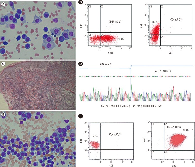
Fig. 1 Morphological features, flow cytometric analysis, immunohistochemical stain, and genetic study of the two cases of CD4+/CD56+AML. (A) Plasmoblast-like neoplastic cells in the first case (Wright Giemsa stain, 400×, Bone marrow); (B) Immunophenotyping features with CD56 and CD4 coexpression in the first case; (C) Skin biopsy showing diffuse infiltration of medium- to large-sized agranular blastic cells into the dermis in the first case (Hematoxylin and Eosin stain, 100×, Skin lesion); (D) The MLL-MLLT10 rearrangement confirmed by direct sequencing in the first case; (E) Plasmacytoid cells in the second case (Wright Giemsa stain; ×400, Bone marrow); and (F) The immunophenotyping features with CD4 and CD56 coexpression in the second case.
Reference
-
1. Garnache-Ottou F, Feuillard J, Saas P. Plasmacytoid dendritic cell leukaemia/lymphoma: towards a well defined entity? Br J Haematol. 2007; 136:539–548. PMID: 17367408.2. Swerdlow SH, Campo E. . WHO classification of tumours of haematopoietic and lymphoid tissues. France: IARC Press;2008. p. 145–147.3. Leung R, Chow EE, Au WY, Chow C, Kwong YL, Lin SY, et al. CD4+/CD56+ hematologic malignancy with rearranged MLL gene. Hum Pathol. 2006; 37:247–249. PMID: 16426929.4. Toya T, Nishimoto N, Koya J, Nakagawa M, Nakamura F, Kandabashi K, et al. The first case of blastic plasmacytoid dendritic cell neoplasm with MLL-ENL rearrangement. Leuk Res. 2012; 36:117–118. PMID: 21831428.5. Yang N, Huh J, Chung WS, Cho MS, Ryu KH, Chung HS. KMT2A (MLL)-MLLT1 rearrangement in blastic plasmacytoid dendritic cell neoplasm. Cancer Genet. 2015; 208:464–467. PMID: 26164398.6. Muñoz L, Nomdedéu JF, Villamor N, Guardia R, Colomer D, Ribera JM, et al. Acute myeloid leukemia with MLL rearrangements: clinicobiological features, prognostic impact and value of flow cytometry in the detection of residual leukemic cells. Leukemia. 2003; 17:76–82. PMID: 12529663.7. Rush PS, Bennett DD, Yang DT. Hematopathology HP 15-5. ASCP case reports. 2015; HP 15-5:1–20.8. Bekkenk MW, Jansen PM, Meijer CJ, Willemze R. CD56+ hematological neoplasms presenting in the skin: a retrospective analysis of 23 new cases and 130 cases from the literature. Ann Oncol. 2004; 15:1097–1108. PMID: 15205205.9. Herling M, Jones D. CD4+/CD56+ hematodermic tumor: the features of an evolving entity and its relationship to dendritic cells. Am J Clin Pathol. 2007; 127:687–700. PMID: 17439829.
- Full Text Links
-
- Actions
-
Cited
- CITED
-
- Close
- Share
-
- Similar articles
-
- A Case of Blastic Plasmacytoid Dendritic Cell Neoplasm in Child
- Plasmacytoid dendritic cell neoplasms
- A Case of Blastic Plasmacytoid Dendritic Cell Neoplasm with Mutations in DNMT3A, TET2, SRSF2, and ATRX Genes
- A Woman with Blastic Plasmacytoid Dendritic Cell Neoplasm
- Blastic Plasmacytoid Dendritic Cell Neoplasm Mimicking Traumatic Hematoma: A Case Report
