Korean J Ophthalmol.
2017 Apr;31(2):165-171. 10.3341/kjo.2017.31.2.165.
A Prospective Study of Anterior Segment Ocular Parameters in Anisometropia
- Affiliations
-
- 1Department of Ophthalmology, University College of Medical Sciences and Guru Teg Bahadur Hospital, Delhi, India. nhsngh.89@gmail.com
- 2Dr. Rajendra Prasad Centre for Ophthalmic Sciences, All India Institute of Medical Sciences, New Delhi, India.
- KMID: 2373474
- DOI: http://doi.org/10.3341/kjo.2017.31.2.165
Abstract
- PURPOSE
The aim of this study was to investigate the differences in anterior segment ocular parameters in anisometropia >1 D.
METHODS
This study included 202 eyes of 101 subjects ranging from 10 to 40 years of age with anisometropia of 1 D or more. The subjects were divided into groups according to anisomyopia, anisoastigmatism, and anisohypermetropia. After providing informed consent, each patient underwent a detailed ophthalmological examination including cycloplegic refraction, best-corrected visual acuity, cover test, axial length (AL) measurement using A-scan ultrasound biometer, keratometry, anterior chamber depth, and central corneal thickness measurement. For each participant, the eye with greater refractive error was compared to the fellow eye via paired t-tests. Correlations between parameters were studied using the Pearson correlation coefficient.
RESULTS
The average age of subjects was 21.7 ± 9.3 years. Of 101 subjects, 31 had anisomyopia; 42 had anisohypermetropia; and 28 had anisoastigmatism. A predisposition toward greater myopia in right eyes was noted in anisomyopia (24 of 31 subjects, 77%). The inter-ocular acuity difference was significant in all three groups (p < 0.01). As the degree of anisometropia increased, there was significant positive correlation in the difference in AL in myopes (r = 0.863, p < 0.01) and hypermetropes (r = 0.669, p < 0.01) and the difference in corneal curvature in anisoastigmatism (r = 0.564, p = 0.002) and hypermetropes (r = 0.376, p = 0.014). A significant positive correlation was also present between the anterior chamber depth difference and refractive difference in hypermetropes (r = 0.359, p = 0.020).
CONCLUSIONS
This study showed that anisomyopia is correlated only with anterior chamber differences. Anisohypermetropia is correlated with AL differences as well as corneal curvature difference and anterior chamber depth difference. The amount of anisoastigmatism correlates only with corneal curvature difference.
MeSH Terms
Figure
-
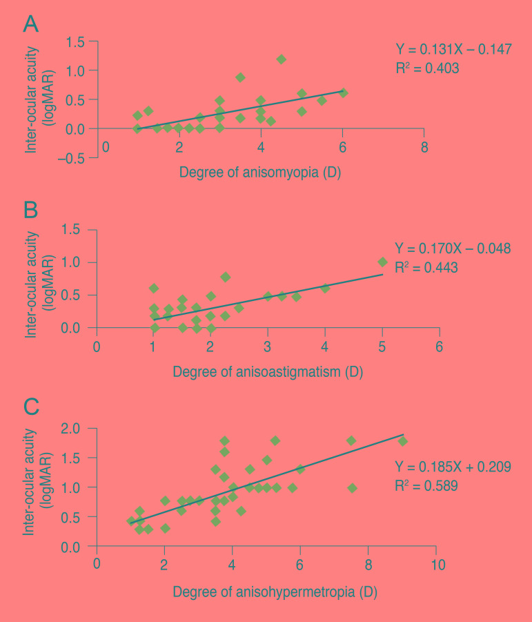
Fig. 1 (A) The correlation between the degree of anisomyopia and inter-ocular acuity difference (IAD). (B) The correlation between anisoastigmatism and IAD. (C) The correlation between anisohypermetropia and IAD. logMAR = logarithm of the minimal angle of resolution; D = diopter.
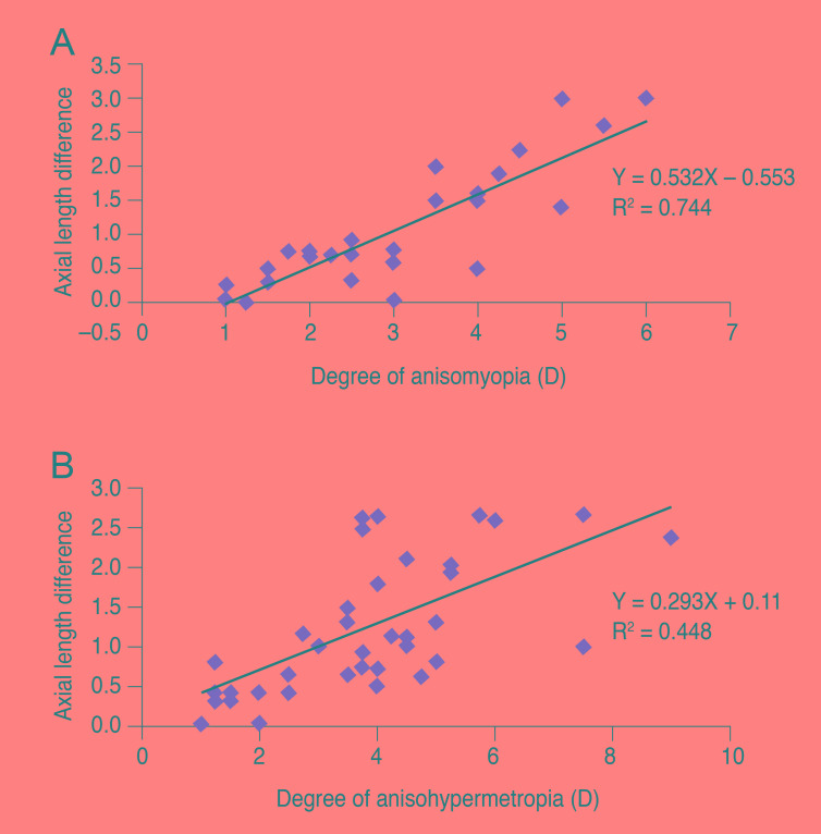
Fig. 2 The linear correlation between the difference in axial length and degree of anisometropia between fellow eyes anisomyopia (A) and anisohypermetropia (B).
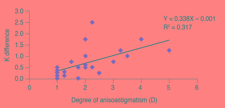
Fig. 3 The correlation between corneal curvature (K) difference and refractive difference between fellow eyes in anisoastigmatism.
Reference
-
1. Gawecki M, Adamski J. Anisometropia and stereopsis. Klin Oczna. 2004; 106:561–563. PMID: 15646468.2. Weakley DR Jr. The association between nonstrabismic anisometropia, amblyopia, and subnormal binocularity. Ophthalmology. 2001; 108:163–171. PMID: 11150283.
Article3. Weakley DR Jr, Birch E, Kip K. The role of anisometropia in the development of accommodative esotropia. J AAPOS. 2001; 5:153–157. PMID: 11404741.
Article4. O'Donoghue L, McClelland JF, Logan NS, et al. Profile of anisometropia and aniso-astigmatism in children: prevalence and association with age, ocular biometric measures, and refractive status. Invest Ophthalmol Vis Sci. 2013; 54:602–608. PMID: 23233258.5. Huynh SC, Wang XY, Ip J, et al. Prevalence and associations of anisometropia and aniso-astigmatism in a population based sample of 6 year old children. Br J Ophthalmol. 2006; 90:597–601. PMID: 16622090.
Article6. Vincent SJ, Collins MJ, Read SA, Carney LG. Myopic anisometropia: ocular characteristics and aetiological considerations. Clin Exp Optom. 2014; 97:291–307. PMID: 24939167.
Article7. Randleman JB. Post-laser in-situ keratomileusis ectasia: current understanding and future directions. Curr Opin Ophthalmol. 2006; 17:406–412. PMID: 16900036.
Article8. Devereux JG, Foster PJ, Baasanhu J, et al. Anterior chamber depth measurement as a screening tool for primary angle-closure glaucoma in an East Asian population. Arch Ophthalmol. 2000; 118:257–263. PMID: 10676792.
Article9. Congdon NG, Youlin Q, Quigley H, et al. Biometry and primary angle-closure glaucoma among Chinese, white, and black populations. Ophthalmology. 1997; 104:1489–1495. PMID: 9307646.
Article10. Olsen T. Sources of error in intraocular lens power calculation. J Cataract Refract Surg. 1992; 18:125–129. PMID: 1564648.
Article11. Holladay JT. Standardizing constants for ultrasonic biometry, keratometry, and intraocular lens power calculations. J Cataract Refract Surg. 1997; 23:1356–1370. PMID: 9423908.
Article12. Benjamin WJ, Borish IM, editors. Borish's clinical refraction. Philadelphia: W. B. Saunders;1998. p. 2–17.13. Troilo D. Neonatal eye growth and emmetropisation: a literature review. Eye (Lond). 1992; 6(Pt 2):154–160. PMID: 1624037.14. Olsen T, Arnarsson A, Sasaki H, et al. On the ocular refractive components: the Reykjavik Eye Study. Acta Ophthalmol Scand. 2007; 85:361–366. PMID: 17286626.
Article15. Wong TY, Foster PJ, Ng TP, et al. Variations in ocular biometry in an adult Chinese population in Singapore: the Tanjong Pagar Survey. Invest Ophthalmol Vis Sci. 2001; 42:73–80. PMID: 11133850.16. Goldschmidt E, Lyhne N, Lam CS. Ocular anisometropia and laterality. Acta Ophthalmol Scand. 2004; 82:175–178. PMID: 15043536.
Article17. Linke SJ, Druchkiv V, Steinberg J, et al. Eye laterality: a comprehensive analysis in refractive surgery candidates. Acta Ophthalmol. 2013; 91:e363–e368. PMID: 23387503.
Article18. Weakley DR. The association between anisometropia, amblyopia, and binocularity in the absence of strabismus. Trans Am Ophthalmol Soc. 1999; 97:987–1021. PMID: 10703148.19. Dobson V, Miller JM, Clifford-Donaldson CE, Harvey EM. Associations between anisometropia, amblyopia, and reduced stereoacuity in a school-aged population with a high prevalence of astigmatism. Invest Ophthalmol Vis Sci. 2008; 49:4427–4436. PMID: 18539935.
Article20. Levi DM, McKee SP, Movshon JA. Visual deficits in anisometropia. Vision Res. 2011; 51:48–57. PMID: 20932989.
Article21. Chen BB, Song FW, Sun ZH, Yang Y. Anisometropia magnitude and visual deficits in previously untreated anisometropic amblyopia. Int J Ophthalmol. 2013; 6:606–610. PMID: 24195034.22. Tian Y, Tarrant J, Wildsoet CF. Optical and biometric characteristics of anisomyopia in human adults. Ophthalmic Physiol Opt. 2011; 31:540–549. PMID: 21797915.
Article23. Curtin BJ, editor. The myopias: basic science and clinical management. Philadelphia: Harper and Row;1985. p. 277–385.24. Tekin K, Cankurtaran V, Inanc M, et al. Effect of myopic anisometropia on anterior and posterior ocular segment parameters. Int Ophthalmol. 2017; 37:377–384. PMID: 27262559.
Article25. Kim SY, Cho SY, Yang JW, et al. The correlation of differences in the ocular component values with the degree of myopic anisometropia. Korean J Ophthalmol. 2013; 27:44–47. PMID: 23372379.
Article26. Logan NS, Gilmartin B, Wildsoet CF, Dunne MC. Posterior retinal contour in adult human anisomyopia. Invest Ophthalmol Vis Sci. 2004; 45:2152–2162. PMID: 15223789.
Article27. Goss DA, Erickson P. Meridional corneal components of myopia progression in young adults and children. Am J Optom Physiol Opt. 1987; 64:475–481. PMID: 3631204.
Article28. Shih YF, Chiang TH, Lin LL. Lens thickness changes among schoolchildren in Taiwan. Invest Ophthalmol Vis Sci. 2009; 50:2637–2644. PMID: 19234352.
Article29. Strang NC, Schmid KL, Carney LG. Hyperopia is predominantly axial in nature. Curr Eye Res. 1998; 17:380–383. PMID: 9561829.
Article30. Demircan S, Gokce G, Yuvaci I, et al. The assessment of anterior and posterior ocular structures in hyperopic anisometropic amblyopia. Med Sci Monit. 2015; 21:1181–1188. PMID: 25910432.
Article31. Yuksel N, Yuksel E, Ozer MD. Evaluation of anterior segment parameters using the Pentacam in hyperopic anisometropic amblyopic and normal eyes. J AAPOS. 2014; 18:248–250. PMID: 24924277.
Article32. Pediatric Eye Disease Investigator Group. The clinical profile of moderate amblyopia in children younger than 7 years. Arch Ophthalmol. 2002; 120:281–287. PMID: 11879130.
- Full Text Links
-
- Actions
-
Cited
- CITED
-
- Close
- Share
-
- Similar articles
-
- The Association Between Amblyopia and Anisometropia in Intermittent Exotropia
- Ocular Myasthenia Gravis in Monozygotic Twins with Mirror-image Myopic Anisometropia
- Department of Ophthalmology, Taegu Fatima Hospital
- The Correlation of Differences in the Ocular Component Values with the Degree of Myopic Anisometropia
- A Longitudinal Change of Spherical Equivalent in Anisometropic Children
