Korean J Ophthalmol.
2017 Apr;31(2):151-158. 10.3341/kjo.2017.31.2.151.
Choroidal Thickness Variation According to Refractive Error Measured by Spectral Domain-optical Coherence Tomography in Korean Children
- Affiliations
-
- 1Cheil Eye Hospital, Daegu, Korea. eyejholee@hotmail.com
- KMID: 2373472
- DOI: http://doi.org/10.3341/kjo.2017.31.2.151
Abstract
- PURPOSE
To assess choroidal thickness (CT) variation according to refractive errors using enhanced-depth imaging optical coherence tomography.
METHODS
Eighty-nine eyes (in 89 children) <±6 diopter were categorized into three groups: hyperopia, emmetropia, and myopia, according to refractive error, and underwent choroidal scans using enhanced-depth imaging-optical coherence tomography. CT was measured at the fovea and at 1 mm and 3 mm nasal (N1 and N3), temporal (T1 and T3), superior (S1 and S3), and inferior (I1 and I3) from the fovea.
RESULTS
Mean foveal CTs were 346.86 µm, 301.97 µm, and 267.46 µm in the hyperopia, emmetropia, and myopia groups, respectively (p < 0.05). CTs at N3 and T3 were 214.59 µm and 318.68 µm, 163.92 µm and 320.79 µm, and 153.93 µm and 295.61 µm in the hyperopia, emmetropia, and myopia groups, respectively (p < 0.05). All CTs in the hyperopia group were thicker than those of other groups (p < 0.05). Fovea was thickest and was significantly thicker than at N3 and I3 in hyperopia (p < 0.05). T3 thickness in the emmetropia and myopia groups was greater than thickness at other areas, particularly the nasal and inferior choroids (p < 0.05). CT was positively correlated with spherical equivalent (p = 0.029).
CONCLUSIONS
In Korean children, CTs were greater in the hyperopia group than in the emmetropia and myopia groups. The temporal choroid was thicker than the nasal choroid, regardless of the refractive error. The thickest location in the hyperopia group was the fovea; however, the temporal choroid was thickest in the emmetropia and myopia groups.
Keyword
MeSH Terms
Figure
-
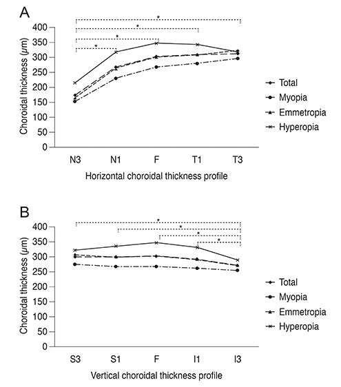
Fig. 1 Horizontal (A) and vertical (B) choroidal thickness profiles. (A) N3–N1, p < 0.001; N3–F, p < 0.001; N3–T1, p < 0.001; N3–T3, p < 0.001. (B) S3–I3, p = 0.001; S1–I3, p = 0.003; F–I3, p = 0.001; I1–I3, p = 0.014. N3 = 3 mm nasal to fovea; N1 = 1 mm nasal to fovea; F = fovea; T1 = 1 mm temporal to fovea; T3 = 3 mm temporal to fovea; S3 = 3 mm superior to fovea; S1 = 1 mm superior to fovea; I1 = 1 mm inferior to fovea; I3 = 3 mm inferior to the fovea. *p-value <0.05 using post-hoc analysis with least significant difference test in all eyes.
Reference
-
1. Nickla DL, Wallman J. The multifunctional choroid. Prog Retin Eye Res. 2010; 29:144–168.2. Povazay B, Hermann B, Unterhuber A, et al. Three-dimensional optical coherence tomography at 1050 nm versus 800 nm in retinal pathologies: enhanced performance and choroidal penetration in cataract patients. J Biomed Opt. 2007; 12:041211.3. Spaide RF, Koizumi H, Pozzoni MC. Enhanced depth imaging spectral-domain optical coherence tomography. Am J Ophthalmol. 2008; 146:496–500.4. Margolis R, Spaide RF. A pilot study of enhanced depth imaging optical coherence tomography of the choroid in normal eyes. Am J Ophthalmol. 2009; 147:811–815.5. Spaide RF. Age-related choroidal atrophy. Am J Ophthalmol. 2009; 147:801–810.6. Torres VL, Brugnoni N, Kaiser PK, Singh AD. Optical coherence tomography enhanced depth imaging of choroidal tumors. Am J Ophthalmol. 2011; 151:586–593.e2.7. Ikuno Y, Kawaguchi K, Nouchi T, Yasuno Y. Choroidal thickness in healthy Japanese subjects. Invest Ophthalmol Vis Sci. 2010; 51:2173–2176.8. Wei WB, Xu L, Jonas JB, et al. Subfoveal choroidal thickness: the Beijing Eye Study. Ophthalmology. 2013; 120:175–180.9. Tan CS, Ouyang Y, Ruiz H, Sadda SR. Diurnal variation of choroidal thickness in normal, healthy subjects measured by spectral domain optical coherence tomography. Invest Ophthalmol Vis Sci. 2012; 53:261–266.10. Read SA, Collins MJ, Vincent SJ, Alonso-Caneiro D. Choroidal thickness in myopic and nonmyopic children assessed with enhanced depth imaging optical coherence tomography. Invest Ophthalmol Vis Sci. 2013; 54:7578–7586.11. Park KA, Oh SY. Choroidal thickness in healthy children. Retina. 2013; 33:1971–1976.12. Hung LF, Wallman J, Smith EL 3rd. Vision-dependent changes in the choroidal thickness of macaque monkeys. Invest Ophthalmol Vis Sci. 2000; 41:1259–1269.13. Troilo D, Nickla DL, Wildsoet CF. Choroidal thickness changes during altered eye growth and refractive state in a primate. Invest Ophthalmol Vis Sci. 2000; 41:1249–1258.14. Zhu X, Wallman J. Temporal properties of compensation for positive and negative spectacle lenses in chicks. Invest Ophthalmol Vis Sci. 2009; 50:37–46.15. Nishi T, Ueda T, Hasegawa T, et al. Choroidal thickness in children with hyperopic anisometropic amblyopia. Br J Ophthalmol. 2014; 98:228–232.16. Demircan A, Altan C, Osmanbasoglu OA, et al. Subfoveal choroidal thickness measurements with enhanced depth imaging optical coherence tomography in patients with nanophthalmos. Br J Ophthalmol. 2014; 98:345–349.17. Yamani A, Wood I, Sugino I, et al. Abnormal collagen fibrils in nanophthalmos: a clinical and histologic study. Am J Ophthalmol. 1999; 127:106–108.18. Oner V, Bulut A, Buyuktarakci S, Kaim M. Influence of hyperopia and amblyopia on choroidal thickness in children. Eur J Ophthalmol. 2016; 26:623–626.19. Read SA, Collins MJ, Vincent SJ, Alonso-Caneiro D. Choroidal thickness in childhood. Invest Ophthalmol Vis Sci. 2013; 54:3586–3593.20. Ruiz-Moreno JM, Flores-Moreno I, Lugo F, et al. Macular choroidal thickness in normal pediatric population measured by swept-source optical coherence tomography. Invest Ophthalmol Vis Sci. 2013; 54:353–359.21. Bill A, Sperber G, Ujiie K. Physiology of the choroidal vascular bed. Int Ophthalmol. 1983; 6:101–107.22. Li XQ, Jeppesen P, Larsen M, Munch IC. Subfoveal choroidal thickness in 1323 children aged 11 to 12 years and association with puberty: the Copenhagen Child Cohort 2000 Eye Study. Invest Ophthalmol Vis Sci. 2014; 55:550–555.
- Full Text Links
-
- Actions
-
Cited
- CITED
-
- Close
- Share
-
- Similar articles
-
- Measurement of Choroidal Thickness in Normal Eyes Using 3D OCT-1000 Spectral Domain Optical Coherence Tomography
- The Relationship among Refractive Power, Axial Length and Choroidal Thickness Measured by SD-OCT in Myopia
- The Variation of Choroidal Thickness and Refractive Error after Cataract Surgery
- Choroidal Thickness at the Outside of Fovea in Diabetic Retinopathy Using Spectral-Domain Optical Coherence Tomography
- Use of Spectral-Domain Optical Coherence Tomography to Analyze Macular Thickness According to Refractive Error
