A Case of Small Bowel GIST Initially Suspected as Peritoneal Seeding of Gastric Cancer
- Affiliations
-
- 1Department of Surgery, Kyung Hee University School of Medicine, Seoul, Korea. kyjho@khmc.or.kr
Abstract
- Gastrointestinal stromal tumors (GISTs) constitute the most common primary mesenchymal tumors of the digestive tract and characteristically express c-kit (CD117). GISTs are the most common non-epithelial tumor of the GI tract and frequently originate from the stomach and small bowel. Specifically, the synchronous occurrence of a GIST with other epithelial tumors is rarely reported. Recently, we discovered one case of a concurrent gastric cancer and a small bowel GIST that was initially suspected to be peritoneal seeding from gastric cancer. The patient was initially admitted with epigastric pain. Gastric cancer with peritoneal seeding was suspected after an evaluation. Following a laparoscopic examination, a distal gastrectomy with D2 lymph node dissection and small-intestine segmental resection was performed. The final pathologic diagnosis was early gastric cancer and high-risk small bowel GIST. The patient refused adjuvant therapy for the GIST, and currently shows no other marked indisposition. He has been disease-free for 14 months.
MeSH Terms
Figure
-

Fig. 1 Gastroscopic finding showed a Bormann type III advanced gastric cancer in the anterior wall of the antrum.
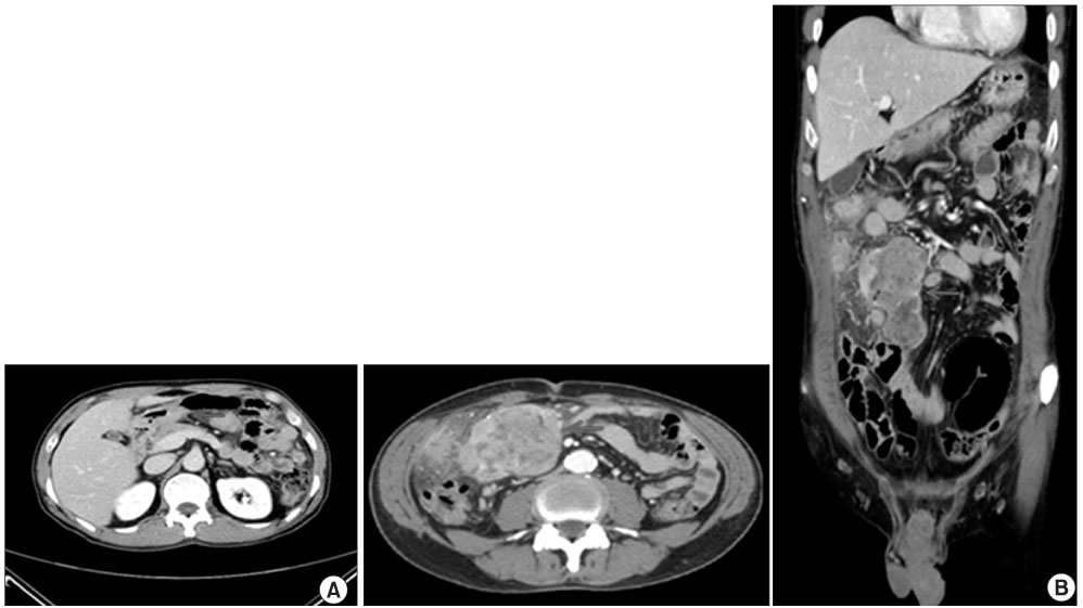
Fig. 2 Abdominal computed tomo graphy suggestive of advanced gastric cancer in the antrum (A) with omental and mesenteric metastatic masses (B).

Fig. 3 (A) In the antrum of the stomach found, an ulcerating lesion. It measured 2×1.5 cm in size and is invaded the submucosa layer. (B) The serosal surface of the small bowel lesion showed a huge multi-lobulating mass, measuring 8×7×7 cm.

Fig. 4 (A) H&E staining of the gastric lesion showed poorly differentiated adenocarcinoma with signet ring cells (×200). (B) H&E staining of the small intestine lesion showed a well-circumscribed tumor tissue, consisted of interlacing or whorling of spindle cells with intervening hyalinized stroma (×200). It showed increased cellularity and frequent mitosis (more than 8/50 HPFs). (C) Immunohistochemical staining of the small bowel mass was positive for CD117.
Reference
-
1. Miettinen M, El-Rifai W, H L, Lasota J. Evaluation of malignancy and prognosis of gastrointestinal stromal tumors: a review. Hum Pathol. 2002. 33:478–483.
Article2. Fletcher CD, Berman JJ, Corless C, Gorstein F, Lasota J, Longley BJ, et al. Diagnosis of gastrointestinal stromal tumors: a consensus approach. Hum Pathol. 2002. 33:459–465.
Article3. Corless CL, Fletcher JA, Heinrich MC. Biology of gastrointestinal stromal tumors. J Clin Oncol. 2004. 22:3813–3825.
Article4. Ozgun YM, Ergul E, Sisman IC, Kusdemir A. Gastric adenocarcinoma and GIST (collision tumors) of the stomach presenting with perforation; first report. Bratisl Lek Listy. 2009. 110:504–505.5. Villias C, Gourgiotis S, Veloudis G, Sampaziotis D, Moreas H. Synchronous early gastric cancer and gastrointestinal stromal tumor in the stomach of a patient with idiopathic thrombocytopenic purpura. J Dig Dis. 2008. 9:104–107.
Article6. O'Sullivan MJ. Gastrointestinal stromal tumors. Pediatr Surg Int. 2009. 25:841–850.7. Lin YL, Tzeng JE, Wei CK, Lin CW. Small gastrointestinal stromal tumor concomitant with early gastric cancer: a case report. World J Gastroenterol. 2006. 12:815–817.
Article8. Maiorana A, Fante R, Maria Cesinaro A, Adriana Fano R. Synchronous occurrence of epithelial and stromal tumors in the stomach: a report of 6 cases. Arch Pathol Lab Med. 2000. 124:682–686.9. Andea AA, Lucas C, Cheng JD, Adsay NV. Synchronous occurrence of epithelial and stromal tumors in the stomach. Arch Pathol Lab Med. 2001. 125:318–319.
Article
- Full Text Links
-
- Actions
-
Cited
- CITED
-
- Close
- Share
-
- Similar articles
-
- Clinical Outcomes according to Primary Treatment in Gastric Cancer Patients with Peritoneal Seeding
- Effect of Radical Removal of Primary and Metastatic Lesions in Gastric Cancer with Peritoneal Seeding
- The Result of Conversion Surgery in Gastric Cancer Patients with Peritoneal Seeding
- A case of superior mesenteric artery syndrome due to peritoneal seeding in a colon cancer patient
- A case of successful surgical treatment for peritoneal seeding of hepatocellular carcinoma after radiotherapy and atezolizumab plus bevacizumab combination treatment
