Treatment of Intracranial Aneurysms with Flow Re-direction Endoluminal Device - A Single Centre Experience with Short-term Follow-up Results
- Affiliations
-
- 1Department of Radiology, Tuen Mun Hospital, Hong Kong.
- 2Department of Radiology, Princess Margaret Hospital, Hong Kong. neeraj.mahboobani@gmail.com
- 3Department of Neurosurgery, Tuen Mun Hospital, Hong Kong.
- KMID: 2371754
- DOI: http://doi.org/10.5469/neuroint.2017.12.1.11
Abstract
- PURPOSE
A flow diverter (FD) is an effective treatment option for intracranial aneurysms. The Flow Re-direction Endoluminal Device (FRED) is a relatively new flow diverter with a unique dual-layer design. We report our experience and short-term results with the FRED.
MATERIALS AND METHODS
We did a retrospective review of all consecutive cases in which the FRED was used to treat intracranial aneurysms at a single institution from March 2014 till December 2015. Clinical parameters, aneurysm characteristics, technical results and short-term outcomes were reviewed.
RESULTS
Eleven intracranial aneurysms were treated with the FRED in 11 patients. The technical device deployment success rate was 100%. Immediate reduction in intra-aneurysmal flow after deployment was noted in 10 cases. The aneurysm occlusion rate at 6 months was 75%. There was 1 complication of in-stent thrombosis immediately after deployment. There was no side branch occlusion, delayed aneurysm rupture, stroke, or intraparenchymal haemorrhage. There was no neurological deficit, morbidity, or mortality.
CONCLUSION
The FRED is a new FD. It has shown to be safe and effective in our series. The unique dual-layer design of the device renders it to have technical advantages over other FDs. The 6-month aneurysm occlusion rate and complication profile of FRED are similar to other FDs.
Keyword
MeSH Terms
Figure
-

Fig. 1 The FRED device. Profile view (A) and illustration (B) of the FRED showing dual-layer design. Reproduced with permission from MicroVention, Inc.

Fig. 2 A 53-year-old man with large left paraophthalmic internal carotid artery (ICA) aneurysm (case 9). (A) Lateral view of left ICA angiogram showing large paraophthalmic aneurysm. (B, C) Immediate post-deployment lateral view of left ICA angiogram at arterial (B) and venous (C) phases with layering of contrast seen on venous (C) phase. (D) Post-deployment anteroposterior (AP) view of left common carotid artery (CCA) angiogram showing no opacification of left ICA beyond the vertical part of petrous segment (arrow). (E) AP view of left ICA angiogram after intra-arterial thrombolysis with abciximab showing partial recanalization of left intracranial ICA. (F) AP view of right ICA angiogram showing crossflow to left anterior (ACA) and middle (MCA) cerebral arteries via anterior communicating artery. (G) CT angiogram of Circle of Willis acquired 2 weeks after FRED deployment showing occluded left ICA (arrow). (H) CT angiogram of Circle of Willis acquired 2 weeks after FRED deployment showing thrombosed aneurysm (arrow) and patent left ACA and MCA. (I) Non-contrast CT brain done prior to CT angiogram of Circle of Willis with image at level of lentiform nuclei - no infarct is seen.

Fig. 3 A 38-year-old man with large ruptured left paraophthalmic internal carotid artery (ICA) aneurysm (case 7). (A) Lateral view of left ICA angiogram showing large paraophthalmic aneurysm. (B, C) Angiogram after deployment of the first FRED showing opacification of aneurysm (B) with contrast washout (C) in sync with ICA indicative of rapid flow within aneurysm. (D, E) Angiogram after deployment of a second FRED in an overlapping manner showing opacification of aneurysm (D) with contrast stasis and layering (E). (F) 6-month follow-up angiogram with illustrated working length (arrowheads) and total length (arrows) of overlapping FREDs. The aneurysm is occluded. Left ophthalmic artery is patent.
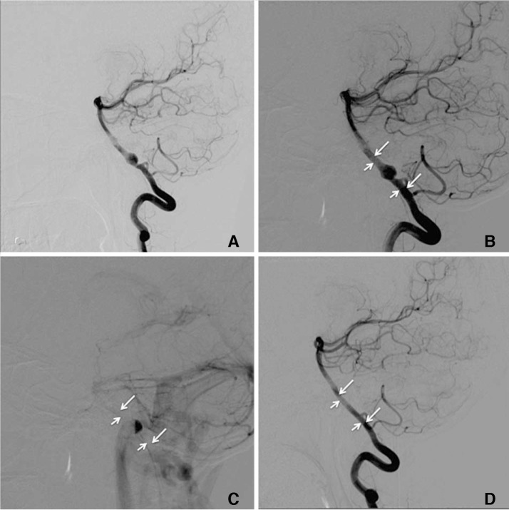
Fig. 4 A 42-year-old woman with dissecting fusiform aneurysm in V4 segment of right vertebral artery (VA; case 4). (A) Lateral view of right VA angiogram showing the aneurysm shortly distal to origin of right posterior inferior cerebellar artery (PICA). (B) Arterial phase of immediate post-FRED deployment angiogram with illustrated working length (short arrows) and total length (long arrows) of the FRED. (C) Venous phase of immediate post-deployment angiogram showing layering of contrast within the aneurysm. (D) 6-month follow-up angiogram showing occluded aneurysm and patent right PICA.
Cited by 1 articles
-
The Evolution of Flow-Diverting Stents for Cerebral Aneurysms; Historical Review, Modern Application, Complications, and Future Direction
Dong-Seong Shin, Christopher P. Carroll, Mohammed Elghareeb, Brian L. Hoh, Bum-Tae Kim
J Korean Neurosurg Soc. 2020;63(2):137-152. doi: 10.3340/jkns.2020.0034.
Reference
-
1. Brinjikji W, Murad MH, Lanzino G, Cloft HJ, Kallmes DF. Endovascular treatment of intracranial aneurysms with flow diverters: a meta-analysis. Stroke. 2013; 44:442–447. PMID: 23321438.
Article2. Alderazi YJ, Shastri D, Kass-Hout T, Prestigiacomo CJ, Gandhi CD. Flow diverters for intracranial aneurysms. Stroke Res Treat. 2014; 2014:415653. PMID: 24967131.
Article3. Zanaty M, Chalouhi N, Tjoumakaris SI, Rosenwasser RH, Gonzalez LF, Jabbour P. Flow-diversion panacea or poison? Front Neurol. 2014; 5:21. PMID: 24592254.
Article4. Diaz O, Gist TL, Manjarez G, Orozco F, Almeida R. Treatment of 14 intracranial aneurysms with the FRED system. J Neurointerv Surg. 2014; 6:614–617. PMID: 24062251.
Article5. Möhlenbruch MA, Herweh C, Jestaedt L, Stampfl S, Schonenberger S, Ringleb PA, et al. The FRED flow-diverter stent for intracranial aneurysms: clinical study to assess safety and efficacy. AJNR Am J Neuroradiol. 2015; 36:1155–1161. PMID: 25721079.
Article6. Kocer N, Islak C, Kizilkilic O, Kocak B, Saglam M, Tureci E. Flow Re-direction Endoluminal Device in treatment of cerebral aneurysms: initial experience with short-term follow-up results. J Neurosurg. 2014; 120:1158–1171. PMID: 24628615.
Article7. Poncyljusz W, Sagan L, Safranow K, Rac M. Initial experience with implantation of novel dual layer flow-diverter device FRED. Wideochir Inne Tech Maloinwazyjne. 2013; 8:258–264. PMID: 24130644.
Article8. O'Kelly CJ, Krings T, Fiorella D, Marotta TR. A novel grading scale for the angiographic assessment of intracranial aneurysms treated using flow diverting stents. Interv Neuroradiol. 2010; 16:133–137. PMID: 20642887.9. Joshi MD, O'Kelly CJ, Krings T, Fiorella D, Marotta TR. Observer variability of an angiographic grading scale used for the assessment of intracranial aneurysms treated with flow-diverting stents. AJNR Am J Neuroradiol. 2013; 34:1589–1592. PMID: 23449648.
Article10. Delgado Almandoz JE, Crandall BM, Scholz JM, Fease JL, Anderson RE, Kadkhodayan Y, et al. Pre-procedure P2Y12 reaction units value predicts perioperative thromboembolic and hemorrhagic complications in patients with cerebral aneurysms treated with the Pipeline Embolization Device. J Neurointerv Surg. 2013; 5:iii3–iii10. PMID: 23314576.
Article11. Tan LA, Keigher KM, Munich SA, Moftakhar R, Lopes DK. Thromboembolic complications with Pipeline Embolization Device placement: impact of procedure time, number of stents and pre-procedure P2Y12 reaction unit (PRU) value. J Neurointerv Surg. 2015; 7:217–221. PMID: 24553344.
Article12. Heller RS, Dandamudi V, Lanfranchi M, Malek AM. Effect of antiplatelet therapy on thromboembolism after flow diversion with the pipeline embolization device. J Neurosurg. 2013; 119:1603–1610. PMID: 23971953.
Article13. Szikora I, Berentei Z, Kulcsar Z, Marosfoi M, Vajda ZS, Lee W, et al. Treatment of intracranial aneurysms by functional reconstruction of the parent artery: the budapest experience with the pipeline embolization device. AJNR Am J Neuroradiol. 2010; 31:1139–1147. PMID: 20150304.
Article
- Full Text Links
-
- Actions
-
Cited
- CITED
-
- Close
- Share
-
- Similar articles
-
- Flow Diverter Device for Treatment of Cerebral Aneurysm with Short-Term Follow Up: Two Case Reports
- A Single Flow Re-direction Endoluminal Device for the Treatment of Large and Giant Anterior Circulation Intracranial Aneurysms
- Flow Diverter Treatment Using a Flow Re-Direction Endoluminal Device for Unruptured Intracranial Vertebral Artery Dissecting Aneurysm: Single-Center Case Series and Technical Considerations
- A Case of Migration of Pipeline Embolization Device Causing Rupture during Treatment of an Unruptured Vertebral Artery Dissecting Aneurysm
- Internal Carotid Artery Reconstruction with a “Mega Flow Diverterâ€: First Experience with the 6×50 mm DERIVO Embolization Device
