Quantitative Muscle Ultrasonography in Carpal Tunnel Syndrome
- Affiliations
-
- 1Department of Rehabilitation Medicine, Chungnam National University School of Medicine, Daejeon, Korea. mksohn@cnuh.co.kr
- KMID: 2371337
- DOI: http://doi.org/10.5535/arm.2016.40.6.1048
Abstract
OBJECTIVE
To assess the reliability of quantitative muscle ultrasonography (US) in healthy subjects and to evaluate the correlation between quantitative muscle US findings and electrodiagnostic study results in patients with carpal tunnel syndrome (CTS). The clinical significance of quantitative muscle US in CTS was also assessed.
METHODS
Twenty patients with CTS and 20 age-matched healthy volunteers were recruited. All control and CTS subjects underwent a bilateral median and ulnar nerve conduction study (NCS) and quantitative muscle US. Transverse US images of the abductor pollicis brevis (APB) and abductor digiti minimi (ADM) were obtained to measure muscle cross-sectional area (CSA), thickness, and echo intensity (EI). EI was determined using computer-assisted, grayscale analysis. Inter-rater and intra-rater reliability for quantitative muscle US in control subjects, and differences in muscle thickness, CSA, and EI between the CTS patient and control groups were analyzed. Relationships between quantitative US parameters and electrodiagnostic study results were evaluated.
RESULTS
Quantitative muscle US had high inter-rater and intra-rater reliability in the control group. Muscle thickness and CSA were significantly decreased, and EI was significantly increased in the APB of the CTS group (all p<0.05). EI demonstrated a significant positive correlation with latency of the median motor and sensory NCS in CTS patients (p<0.05).
CONCLUSION
These findings suggest that quantitative muscle US parameters may be useful for detecting muscle changes in CTS. Further study involving patients with other neuromuscular diseases is needed to evaluate peripheral muscle change using quantitative muscle US.
Keyword
MeSH Terms
Figure
-
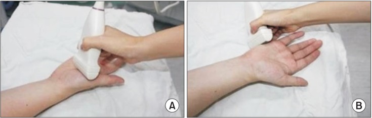
Fig. 1 Position for quantitative muscle ultrasonographic measurements in the right abductor pollicis brevis (A) and abductor digiti minimi (B).
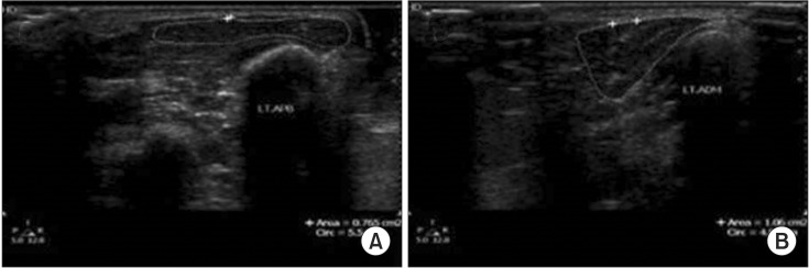
Fig. 2 Transverse ultrasonography images of the left abductor pollicis brevis (APB) and abductor digiti minimi (ADM) of a control subject. Calculation of muscle thickness and cross-sectional area in the APB (A) and ADM (B).
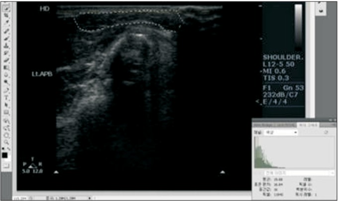
Fig. 3 Echo intensity (EI) analysis of ultrasound images. Grayscale images were used to determine muscle EI within each region of interest (ROI). The mean and standard deviation of the pixel brightness in each ROI were automatically calculated by Photoshop software.
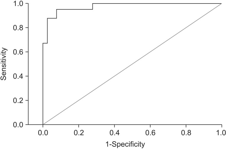
Fig. 4 Receiver operating characteristic (ROC) curve for abductor pollicis brevis muscle echo intensity to distinguish patients with carpal tunnel syndrome from control subjects. The optimal cutoff point was 22.60 (sensitivity 92.5%, specificity 92.5%).
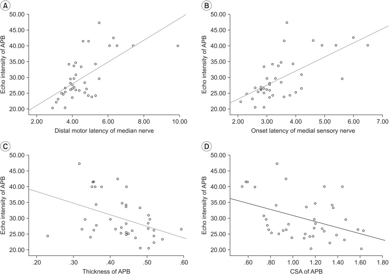
Fig. 5 Pearson correlation scatter plots for study results of echo intensity of abductor pollicis brevis (APB). (A) Distal motor latency of the median nerve (r=0.65). (B) Onset latency of the median sensory nerve (r=0.67). (C) Thickness of APB (r=−0.4). (D) Cross-sectional area (CSA) of APB (r=−0.41).
Reference
-
1. Stevens JC. AAEM minimonograph #26: the electrodiagnosis of carpal tunnel syndrome. Muscle Nerve. 1997; 20:1477–1486. PMID: 9390659.
Article2. Werner RA, Andary M. Electrodiagnostic evaluation of carpal tunnel syndrome. Muscle Nerve. 2011; 44:597–607. PMID: 21922474.
Article3. Al-Shekhlee A, Shapiro BE, Preston DC. Iatrogenic complications and risks of nerve conduction studies and needle electromyography. Muscle Nerve. 2003; 27:517–526. PMID: 12707972.
Article4. Rubin DI. Technical issues and potential complications of nerve conduction studies and needle electromyography. Neurol Clin. 2012; 30:685–710. PMID: 22361380.
Article5. Buchberger W, Judmaier W, Birbamer G, Lener M, Schmidauer C. Carpal tunnel syndrome: diagnosis with high-resolution sonography. AJR Am J Roentgenol. 1992; 159:793–798. PMID: 1529845.
Article6. Koenig RW, Pedro MT, Heinen CP, Schmidt T, Richter HP, Antoniadis G, et al. High-resolution ultrasonography in evaluating peripheral nerve entrapment and trauma. Neurosurg Focus. 2009; 26:E13.
Article7. Gellhorn AC, Carlson MJ. Inter-rater, intra-rater, and inter-machine reliability of quantitative ultrasound measurements of the patellar tendon. Ultrasound Med Biol. 2013; 39:791–796. PMID: 23465140.
Article8. Reimers CD, Schlotter B, Eicke BM, Witt TN. Calf enlargement in neuromuscular diseases: a quantitative ultrasound study in 350 patients and review of the literature. J Neurol Sci. 1996; 143:46–56. PMID: 8981297.
Article9. Schmidt R, Voit T. Ultrasound measurement of quadriceps muscle in the first year of life: normal values and application to spinal muscular atrophy. Neuropediatrics. 1993; 24:36–42. PMID: 8474609.
Article10. Kamala D, Suresh S, Githa K. Real-time ultrasonography in neuromuscular problems in children. J Clin Ultrasound. 1985; 13:465–468. PMID: 3932477.11. Zuberi SM, Matta N, Nawaz S, Stephenson JB, McWilliam RC, Hollman A. Muscle ultrasound in the assessment of suspected neuromuscular disease in childhood. Neuromuscul Disord. 1999; 9:203–207. PMID: 10399745.
Article12. Pillen S, Tak RO, Zwarts MJ, Lammens MM, Verrijp KN, Arts IM, et al. Skeletal muscle ultrasound: correlation between fibrous tissue and echo intensity. Ultrasound Med Biol. 2009; 35:443–446. PMID: 19081667.
Article13. Arts IM, Pillen S, Schelhaas HJ, Overeem S, Zwarts MJ. Normal values for quantitative muscle ultrasonography in adults. Muscle Nerve. 2010; 41:32–41. PMID: 19722256.
Article14. Maurits NM, Bollen AE, Windhausen A, De Jager AE, Van Der Hoeven JH. Muscle ultrasound analysis: normal values and differentiation between myopathies and neuropathies. Ultrasound Med Biol. 2003; 29:215–225. PMID: 12659909.
Article15. Bargfrede M, Schwennicke A, Tumani H, Reimers CD. Quantitative ultrasonography in focal neuropathies as compared to clinical and EMG findings. Eur J Ultrasound. 1999; 10:21–29. PMID: 10502636.
Article16. Reimers K, Reimers CD, Wagner S, Paetzke I, Pongratz DE. Skeletal muscle sonography: a correlative study of echogenicity and morphology. J Ultrasound Med. 1993; 12:73–77. PMID: 8468739.
Article17. Arts IM, van Rooij FG, Overeem S, Pillen S, Janssen HM, Schelhaas HJ, et al. Quantitative muscle ultrasonography in amyotrophic lateral sclerosis. Ultrasound Med Biol. 2008; 34:354–361. PMID: 17964067.
Article18. de Krom MC, Knipschild PG, Kester AD, Thijs CT, Boekkooi PF, Spaans F. Carpal tunnel syndrome: prevalence in the general population. J Clin Epidemiol. 1992; 45:373–376. PMID: 1569433.
Article19. Kim JS, Seok HY, Kim BJ. The significance of muscle echo intensity on ultrasound for focal neuropathy: the median- to ulnar-innervated muscle echo intensity ratio in carpal tunnel syndrome. Clin Neurophysiol. 2016; 127:880–885. PMID: 25998202.
Article20. Scholten RR, Pillen S, Verrips A, Zwarts MJ. Quantitative ultrasonography of skeletal muscles in children: normal values. Muscle Nerve. 2003; 27:693–698. PMID: 12766980.
Article21. Dumitru D, Amato AA, Zwarts M. Electrodiagnostic medicine. 2nd ed. Philadelphia: Hanley & Belfus;2002. p. 1062.22. Padua L, LoMonaco M, Gregori B, Valente EM, Padua R, Tonali P. Neurophysiological classification and sensitivity in 500 carpal tunnel syndrome hands. Acta Neurol Scand. 1997; 96:211–217. PMID: 9325471.
Article23. Fleiss JL. Design and analysis of clinical experiments. New York: John Wiley & Sons;1999. p. 1–28.24. Schmidt WA, Schmidt H, Schicke B, Gromnica-Ihle E. Standard reference values for musculoskeletal ultrasonography. Ann Rheum Dis. 2004; 63:988–994. PMID: 15249327.
Article25. Beekman R, Visser LH. Sonography in the diagnosis of carpal tunnel syndrome: a critical review of the literature. Muscle Nerve. 2003; 27:26–33. PMID: 12508291.
Article26. Pillen S, Arts IM, Zwarts MJ. Muscle ultrasound in neuromuscular disorders. Muscle Nerve. 2008; 37:679–693. PMID: 18506712.
Article
- Full Text Links
-
- Actions
-
Cited
- CITED
-
- Close
- Share
-
- Similar articles
-
- The Abnormal Ultrasonographic Findings of Carpal Tunnel in Carpal Tunnel Syndrome
- Extraskeletal Chondroma Causing Carpal Tunnel Syndrome: A Case Report
- Ultrasound-Guided Nerve Hydrodissection for Carpal Tunnel Syndrome
- RE: Value of Power Doppler and Gray-Scale US in the Diagnosis of Carpal Tunnel Syndrome: Contribution of Cross-Sectional Area just before the Tunnel Inlet as Compared with the Cross-Sectional Area at the Tunnel
- Carpal Tunnel Syndrome Caused by Anatomic Variation of Flexor Digitorum Superficialis of Little Finger
