J Korean Ophthalmol Soc.
2017 Feb;58(2):226-229. 10.3341/jkos.2017.58.2.226.
A Case of Acute Interstitial Keratitis in a Patient with Acquired Syphilis
- Affiliations
-
- 1Department of Ophthalmology, Chosun University School of Medicine, Gwangju, Korea. ophkoh@hanmail.net
- KMID: 2369474
- DOI: http://doi.org/10.3341/jkos.2017.58.2.226
Abstract
- PURPOSE
To report a case of acute interstitial keratitis as the first clinical sign in a patient with latent syphilis.
CASE SUMMARY
A 23-year-old female presented with visual impairment and discomfort in her right eye that developed 3 days earlier. The visual acuity in the right eye was 20/200 and corrected to 20/100, and slit lamp examination showed round sub-epithelial opacification in the central cornea with stromal edema and neovascularization on the cornea of the right eye. Whole body tests including serological tests were performed. Under the suspicion of acute interstitial keratitis, topical antibiotics and steroids were applied 4 times a day initially. Serological tests were reactive for venereal disease research laboratory test (VDRL). Under the suspicion of acute interstitial keratitis due to syphilis, fluorescent treponemal antibody absorption test IgM/IgG (FTA-ABS IgM/IgG) was performed; a positive result for FTA-ABS IgG led to diagnosis of acute interstitial keratitis with latent syphilis. During treatment, systemic doxycycline 200 mg for 4 weeks with topical antibiotics and steroids were administered, the opacity and edema of the cornea regressed after 2 weeks of treatment, and visual acuity in the patient's right eye improved to 20/20.
CONCLUSIONS
We report an unusual case of acute interstitial keratitis as the first clinical manifestation of latent syphilis in an immunocompetent patient.
MeSH Terms
-
Anti-Bacterial Agents
Cornea
Diagnosis
Doxycycline
Edema
Female
Fluorescent Treponemal Antibody-Absorption Test
Humans
Immunoglobulin G
Keratitis*
Patient Rights
Serologic Tests
Sexually Transmitted Diseases
Slit Lamp
Steroids
Syphilis*
Syphilis, Latent
Treponema pallidum
Vision Disorders
Visual Acuity
Young Adult
Anti-Bacterial Agents
Doxycycline
Immunoglobulin G
Steroids
Figure
-
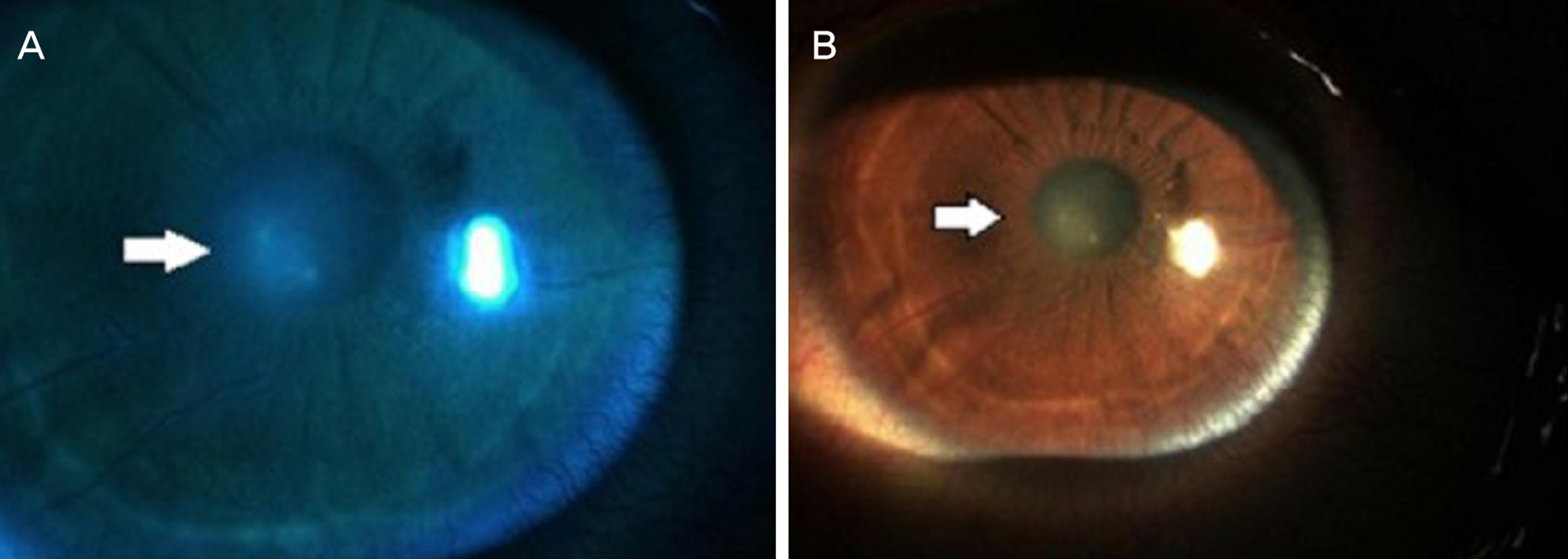
Figure 1. Anterior segmentation photo of deep stromal opacifications, patchy in outline (arrows), and accompanied by the presence of new vessels on cornea of the right eye at first visit. (A) Using blue filter. (B) Without using filter.

Figure 2. Cornea central thickness increased on the right eye (576 μ m). (A) Compared to the left eye (479 μ m), and (B) in anterior segment optical coherence tomography (AOCT) at first visit.

Figure 3. Anterior Segmentation photo of the right eye that the opacity and edema of cornea regressed after 2 weeks (arrows). (A) Without using filter. (B) Using blue filter.
Reference
-
References
1. Kiss S, Damico FM, Young LH. Ocular manifestations and treat-ment of syphilis. Semin Ophthalmol. 2005; 20:161–7.
Article2. Puech C, Gennai S, Pavese P. . Ocular manifestations of syph-ilis: recent cases over a 2.5-year period. Graefes Arch Clin Exp Ophthalmol. 2010; 248:1623–9.
Article3. Smith JL. Testing for congenital syphilis in interstitial keratitis. Am J Ophthalmol. 1971; 72:816–20.
Article4. Lautenschlager S. Diagnosis of syphilis: clinical and laboratory problems. J Dtsch Dermatol Ges. 2006; 4:1058–75.
Article5. Doris JP, Saha K, Jones NP, Sukthankar A. Ocular syphilis: the new epidemic. Eye (Lond). 2006; 20:703–5.
Article6. Gaudio PA. Update on ocular syphilis. Curr Opin Ophthalmol. 2006; 17:562–6.
Article7. Yoon KC, Im SK, Seo MS, Park YG. Neurosyphilitic episcleritis. Acta Ophthalmol Scand. 2005; 83:265–6.
Article8. Aldave AJ, King JA, Cunningham ET. Ocular syphilis. Curr Opin Ophthalmol. 2001; 12:433–41.
Article9. Ratnam S. The laboratory diagnosis of syphilis. Can J Infect Dis Med Microbiol. 2005; 16:45–51.
Article10. Workowski KA, Bolan GA. Sexually transmitted diseases treat-ment guidelines, 2015. MMWR Recomm Rep. 2015; 64((RR-03)):1–137.11. Mannis MJ, Holland EJ. Cornea. 4th ed. Sacramento: Elsivier;2016; 996–1012.
- Full Text Links
-
- Actions
-
Cited
- CITED
-
- Close
- Share
-
- Similar articles
-
- A Case of Acute Interstitial Keratitis with Congenital Syphilis
- A Case of Congenital Syphilitic Interstitial Keratitis
- A Case of Interstitial Keratitis due to Congenital Syphilis
- 3 Cases of Interstitial Keratitis Occurred in Congenital Syphilitic Patients
- 5 Cases of Interstitial Keratitis Due to Congenital Syphilis
