Automatic detection of tooth cracks in optical coherence tomography images
- Affiliations
-
- 1Dental Research Institute, Seoul National University School of Dentistry, Seoul, Korea. wjyi@snu.ac.kr
- 2Department of Biomedical Radiation Sciences, Seoul National University Graduate School of Convergence Science and Technology, Seoul, Korea.
- 3Department of Oral and Maxillofacial Radiology, Seoul National University School of Dentistry, Seoul, Korea.
- KMID: 2368886
- DOI: http://doi.org/10.5051/jpis.2017.47.1.41
Abstract
- PURPOSE
The aims of the present study were to compare the image quality and visibility of tooth cracks between conventional methods and swept-source optical coherence tomography (SS-OCT) and to develop an automatic detection technique for tooth cracks by SS-OCT imaging.
METHODS
We evaluated SS-OCT with a near-infrared wavelength centered at 1,310 nm over a spectral bandwidth of 100 nm at a rate of 50 kHz as a new diagnostic tool for the detection of tooth cracks. The reliability of the SS-OCT images was verified by comparing the crack lines with those detected using conventional methods. After performing preprocessing of the obtained SS-OCT images to emphasize cracks, an algorithm was developed and verified to detect tooth cracks automatically.
RESULTS
The detection capability of SS-OCT was superior or comparable to that of trans-illumination, which did not discriminate among the cracks according to depth. Other conventional methods for the detection of tooth cracks did not sense initial cracks with a width of less than 100 μm. However, SS-OCT detected cracks of all sizes, ranging from craze lines to split teeth, and the crack lines were automatically detected in images using the Hough transform.
CONCLUSIONS
We were able to distinguish structural cracks, craze lines, and split lines in tooth cracks using SS-OCT images, and to automatically detect the position of various cracks in the OCT images. Therefore, the detection capability of SS-OCT images provides a useful diagnostic tool for cracked tooth syndrome.
Keyword
MeSH Terms
Figure
-
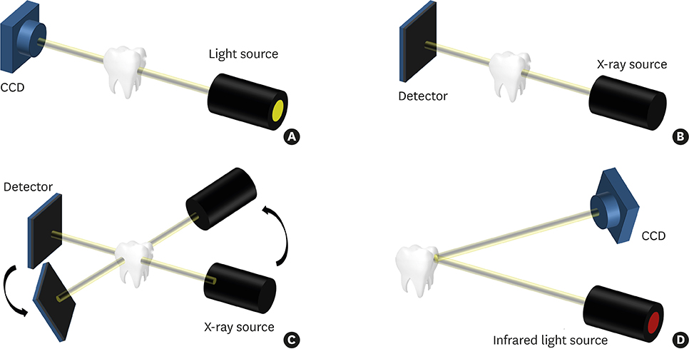
Figure 1 The 4 imaging techniques used in the study. (A) Trans-illumination. A strong light is shone on one side of a tooth and the tooth image is acquired on the other side to identify cracks. (B) An intraoral X-ray is used to acquire a 2-dimensional image of a tooth using radiation. (C) CBCT is used to reconstruct a 3-dimensional image through image processing after obtaining radiographs from various angles. (D) OCT is used to acquire a cross-sectional image of a tooth using an infrared ray of 1,310 nm. CBCT, cone-beam computed tomography; OCT, optical coherence tomography; CCD, charge-coupled detector.
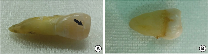
Figure 2 Crack-induced tooth sample. (A) Black arrow indicates the crack. (B) Cracks are difficult to observe with the naked eye.

Figure 3 Results of 4 imaging techniques. The red line indicates the cross-section of CBCT and the OCT scan line. Red and blue arrows indicate crack lines. The blue circle is a false-positive crack line. E, D, and the DEJ are clearly distinguished in the OCT images. (A, E, I) Trans-illumination images. (B, F, J) Intraoral radiography images. (C, G, K) CBCT images. (D, H, L) OCT images. CBCT, cone-beam computed tomography; E, enamel; D, dentine; DEJ, dentine-enamel junction; OCT, optical coherence tomography.

Figure 4 (A, D, G, J, M, and P) Single OCT images showing high noise. (B, E, H, K, N, and Q) OCT images were acquired 100 times, indicating that the noise was improved by image averaging. (C, F, I, L, O, and R) After averaging, crack lines were detected with the Hough transform. OCT, optical coherence tomography.
Reference
-
1. Ehrmann EH, Tyas MJ. Cracked tooth syndrome: diagnosis, treatment and correlation between symptoms and post-extraction findings. Aust Dent J. 1990; 35:105–112.
Article2. Lynch CD, McConnell RJ. The cracked tooth syndrome. J Can Dent Assoc. 2002; 68:470–475.
Article3. Kahler W. The cracked tooth conundrum: terminology, classification, diagnosis, and management. Am J Dent. 2008; 21:275–282.
Article4. Hayashi M, Kinomoto Y, Miura M, Sato I, Takeshige F, Ebisu S. Short-term evaluation of intentional replantation of vertically fractured roots reconstructed with dentin-bonded resin. J Endod. 2002; 28:120–124.
Article5. Lin CC, Tsai YL, Li UM, Chang YC, Lin CP, Jeng JH. Horizontal/oblique root fractures in the palatal root of maxillary molars with associated periodontal destruction: case reports. Int Endod J. 2008; 41:442–447.
Article6. Oztürk M, Unal GC. A successful treatment of vertical root fracture: a case report and 4-year follow-up. Dent Traumatol. 2008; 24:e56–60.
Article7. Liewehr FR. An inexpensive device for transillumination. J Endod. 2001; 27:130–131.
Article8. Mathew S, Thangavel B, Mathew CA, Kailasam S, Kumaravadivel K, Das A. Diagnosis of cracked tooth syndrome. J Pharm Bioallied Sci. 2012; 4:S242–4.
Article9. Whaites E, Drage N. Essentials of dental radiography and radiology. 5th ed. Oxford: Churchill Livingstone;2013.
Article10. Liang X, Jacobs R, Hassan B, Li L, Pauwels R, Corpas L, et al. A comparative evaluation of cone beam computed tomography (CBCT) and multi-slice CT (MSCT) Part I. On subjective image quality. Eur J Radiol. 2010; 75:265–269.
Article11. Parameswaran A. Sturdevant’s art and science of operative dentistry. J Conserv Dent. 2013; 16:480.
Article12. Braz AK, Kyotoku BB, Braz R, Gomes AS. Evaluation of crack propagation in dental composites by optical coherence tomography. Dent Mater. 2009; 25:74–79.13. Nakajima Y, Shimada Y, Miyashin M, Takagi Y, Tagami J, Sumi Y. Noninvasive cross-sectional imaging of incomplete crown fractures (cracks) using swept-source optical coherence tomography. Int Endod J. 2012; 45:933–941.14. Imai K, Shimada Y, Sadr A, Sumi Y, Tagami J. Noninvasive cross-sectional visualization of enamel cracks by optical coherence tomography in vitro . J Endod. 2012; 38:1269–1274.
Article15. Hsieh YS, Ho YC, Lee SY, Chuang CC, Tsai JC, Lin KF, et al. Dental optical coherence tomography. Sensors (Basel). 2013; 13:8928–8949.
Article16. Shimada Y, Sadr A, Sumi Y, Tagami J. Application of optical coherence tomography (oct) for diagnosis of caries, cracks, and defects of restorations. Curr Oral Health Rep. 2015; 2:73–80.
Article17. Lee SH, Lee JJ, Chung HJ, Park JT, Kim HJ. Dental optical coherence tomography: new potential diagnostic system for cracked-tooth syndrome. Surg Radiol Anat. 2016; 38:49–54.
Article18. Huang D, Swanson EA, Lin CP, Schuman JS, Stinson WG, Chang W, et al. Optical coherence tomography. Science. 1991; 254:1178–1181.
Article19. Drexler W, Fujimoto JG. Optical coherence tomography: technology and applications. Berlin: Springer;2008.
Article20. Lee RC, Kang H, Darling CL, Fried D. Automated assessment of the remineralization of artificial enamel lesions with polarization-sensitive optical coherence tomography. Biomed Opt Express. 2014; 5:2950–2962.
Article21. Wada I, Shimada Y, Ikeda M, Sadr A, Nakashima S, Tagami J, et al. Clinical assessment of non carious cervical lesion using swept-source optical coherence tomography. J Biophotonics. 2015; 8:846–854.
Article22. Scarfe WC, Levin MD, Gane D, Farman AG. Use of cone beam computed tomography in endodontics. Int J Dent. 2009; 2009:634567.
Article23. Clark DJ, Sheets CG, Paquette JM. Definitive diagnosis of early enamel and dentin cracks based on microscopic evaluation. J Esthet Restor Dent. 2003; 15:391–401.
Article24. Dummer PM, Kelly T, Meghji A, Sheikh I, Vanitchai JT. An in vitro study of the quality of root fillings in teeth obturated by lateral condensation of gutta-percha or Thermafil obturators. Int Endod J. 1993; 26:99–105.
Article25. Kajan ZD, Taromsari M. Value of cone beam CT in detection of dental root fractures. Dentomaxillofac Radiol. 2012; 41:3–10.
Article
- Full Text Links
-
- Actions
-
Cited
- CITED
-
- Close
- Share
-
- Similar articles
-
- Early caries detection using optical coherence tomography: a review of the literature
- Applications of Optical Imaging System in Dentistry
- Availability of Optical Coherence Tomography in Diagnosis and Classification of Choroidal Neovascularization
- Optical Imaging and Its Clinical Application in Otorhinolaryngology
- Optimization of Percutaneous Coronary Intervention Using Optical Coherence Tomography
