Korean J Ophthalmol.
2017 Feb;31(1):71-79. 10.3341/kjo.2017.31.1.71.
Initial Pattern of Optic Nerve Enhancement in Korean Patients with Unilateral Optic Neuritis
- Affiliations
-
- 1Department of Ophthalmology, Samsung Medical Center, Sungkyunkwan University School of Medicine, Seoul, Korea. syoh@skku.edu
- 2Center for Clinical Specialty, Department of Ophthalmology, National Cancer Center, Goyang, Korea.
- KMID: 2368681
- DOI: http://doi.org/10.3341/kjo.2017.31.1.71
Abstract
- PURPOSE
The purpose of this study was to demonstrate whether the pattern of optic nerve enhancement in magnetic resonance imaging (MRI) can help to differentiate between idiopathic optic neuritis (ON), neuromyelitis optica (NMO), and multiple sclerosis (MS) in unilateral ON.
METHODS
An MRI of the brain and orbits was obtained in patients with acute unilateral ON. Patients with ON were divided into three groups: NMO, MS, and idiopathic ON. The length and location of the abnormal optic nerve enhancement were compared for ON eyes with and without NMO or MS. The correlation between the pattern of optic nerve enhancement and the outcome of visual function was analyzed.
RESULTS
Of the 36 patients with ON who underwent an MRI within 2 weeks of the onset, 19 were diagnosed with idiopathic ON, 9 with NMO, and 8 with MS. Enhancement of the optic nerve occurred in 21 patients (58.3%) and was limited to the orbital segment in 12 patients. Neither the length nor the location of the optic nerve enhancement was significantly correlated with visual functions other than contrast sensitivity or the diagnosis of idiopathic ON, MS, or NMO. Patients with greater extent of optic nerve sheath enhancement and more posterior segment involvement showed higher contrast sensitivity.
CONCLUSIONS
Our data revealed that the pattern of optic nerve enhancement was not associated with diagnosis of idiopathic ON, NMO, or MS in Korean patients with unilateral ON. We believe further studies that include different ethnic groups will lead to a more definitive answer on this subject.
Keyword
MeSH Terms
Figure
-

Fig. 1 Axial views of gadolinium-enhanced T1-weighted fat-suppressed magnetic resonance imaging showing abnormal enhancement of the orbital segments of the left optic nerve.
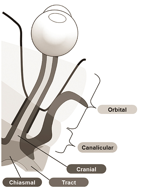
Fig. 2 Illustrations of the segments of the optic nerve used in this study.
Reference
-
1. Rodriguez M, Siva A, Cross SA, et al. Optic neuritis: a population-based study in Olmsted County, Minnesota. Neurology. 1995; 45:244–250.2. Lucchinetti CF, Mandler RN, McGavern D, et al. A role for humoral mechanisms in the pathogenesis of Devic's neuromyelitis optica. Brain. 2002; 125(Pt 7):1450–1461.3. Mandler RN, Davis LE, Jeffery DR, Kornfeld M. Devic's neuromyelitis optica: a clinicopathological study of 8 patients. Ann Neurol. 1993; 34:162–168.4. Lefkowitz D, Angelo JN. Neuromyelitis optica with unusual vascular changes. Arch Neurol. 1984; 41:1103–1105.5. Stansbury FC. Neuromyelitis optica; presentation of five cases, with pathologic study, and review of literature. Arch Ophthal. 1949; 42:292.6. Scott GI. Neuromyelitis optica. Am J Ophthalmol. 1952; 35:755–764.7. Herges K, de Jong BA, Kolkowitz I, et al. Protective effect of an elastase inhibitor in a neuromyelitis optica-like disease driven by a peptide of myelin oligodendroglial glycoprotein. Mult Scler. 2012; 18:398–408.8. Krumbholz M, Faber H, Steinmeyer F, et al. Interferon-beta increases BAFF levels in multiple sclerosis: implications for B cell autoimmunity. Brain. 2008; 131(Pt 6):1455–1463.9. Palace J, Leite MI, Nairne A, Vincent A. Interferon Beta treatment in neuromyelitis optica: increase in relapses and aquaporin 4 antibody titers. Arch Neurol. 2010; 67:1016–1017.10. Tanaka M, Tanaka K, Komori M. Interferon-beta(1b) treatment in neuromyelitis optica. Eur Neurol. 2009; 62:167–170.11. Barnett MH, Prineas JW, Buckland ME, et al. Massive astrocyte destruction in neuromyelitis optica despite natalizumab therapy. Mult Scler. 2012; 18:108–112.12. Kirveskari J, Bono P, Granfors K, et al. Expression of alpha4-integrins on human neutrophils. J Leukoc Biol. 2000; 68:243–250.13. Wingerchuk DM, Lennon VA, Pittock SJ, et al. Revised diagnostic criteria for neuromyelitis optica. Neurology. 2006; 66:1485–1489.14. Beck RW, Gal RL, Bhatti MT, et al. Visual function more than 10 years after optic neuritis: experience of the optic neuritis treatment trial. Am J Ophthalmol. 2004; 137:77–83.15. Pau D, Al Zubidi N, Yalamanchili S, et al. Optic neuritis. Eye (Lond). 2011; 25:833–842.16. Khanna S, Sharma A, Huecker J, et al. Magnetic resonance imaging of optic neuritis in patients with neuromyelitis optica versus multiple sclerosis. J Neuroophthalmol. 2012; 32:216–220.17. Bambach MR, Mitchell RJ. Estimating the human recovery costs of seriously injured road crash casualties. Accid Anal Prev. 2015; 85:177–185.18. Polman CH, Reingold SC, Edan G, et al. Diagnostic criteria for multiple sclerosis: 2005 revisions to the “McDonald Criteria”. Ann Neurol. 2005; 58:840–846.19. Kang ES, Min JH, Lee KH, Kim BJ. Clinical usefulness of cell-based indirect immunofluorescence assay for the detection of aquaporin-4 antibodies in neuromyelitis optica spectrum disorder. Ann Lab Med. 2012; 32:331–338.20. Isayama Y, Takahashi T, Shimoyoma T, Yamadori A. Acute optic neuritis and multiple sclerosis. Neurology. 1982; 32:73–76.21. Lim SA, Goh KY, Tow S, et al. Optic neuritis in Singapore. Singapore Med J. 2008; 49:667–671.22. Lin YC, Yen MY, Hsu WM, et al. Low conversion rate to multiple sclerosis in idiopathic optic neuritis patients in Taiwan. Jpn J Ophthalmol. 2006; 50:170–175.23. Wakakura M, Minei-Higa R, Oono S, et al. Baseline features of idiopathic optic neuritis as determined by a multicenter treatment trial in Japan: Optic Neuritis Treatment Trial Multicenter Cooperative Research Group (ONMRG). Jpn J Ophthalmol. 1999; 43:127–132.24. Wang JC, Tow S, Aung T, et al. The presentation, aetiology, management and outcome of optic neuritis in an Asian population. Clin Exp Ophthalmol. 2001; 29:312–315.25. Beck RW, Arrington J, Murtagh FR, et al. Brain magnetic resonance imaging in acute optic neuritis: Experience of the Optic Neuritis Study Group. Arch Neurol. 1993; 50:841–846.26. Hickman SJ, Miszkiel KA, Plant GT, Miller DH. The optic nerve sheath on MRI in acute optic neuritis. Neuroradiology. 2005; 47:51–55.27. Fazzone HE, Lefton DR, Kupersmith MJ. Optic neuritis: correlation of pain and magnetic resonance imaging. Ophthalmology. 2003; 110:1646–1649.28. Kupersmith MJ, Alban T, Zeiffer B, Lefton D. Contrast-enhanced MRI in acute optic neuritis: relationship to visual performance. Brain. 2002; 125(Pt 4):812–822.29. Lim SA, Sitoh YY, Chng SM, et al. Magnetic resonance imaging in acute optic neuritis in Singapore. Ann Acad Med Singapore. 2009; 38:821–826.30. Wingerchuk DM, Lennon VA, Lucchinetti CF, et al. The spectrum of neuromyelitis optica. Lancet Neurol. 2007; 6:805–815.31. Trobe JD, Beck RW, Moke PS, Cleary PA. Contrast sensitivity and other vision tests in the optic neuritis treatment trial. Am J Ophthalmol. 1996; 121:547–553.32. Park KA, Kim J, Oh SY. Analysis of spectral domain optical coherence tomography measurements in optic neuritis: differences in neuromyelitis optica, multiple sclerosis, isolated optic neuritis and normal healthy controls. Acta Ophthalmol. 2014; 92:e57–e65.
