Effect of different lateral occlusion schemes on peri-implant strain: A laboratory study
- Affiliations
-
- 1Restorative Section, Melbourne Dental School, Melbourne University, Victoria, Australia. jaafar.abduo@unimelb.edu.au
- KMID: 2368211
- DOI: http://doi.org/10.4047/jap.2017.9.1.45
Abstract
- PURPOSE
This study aims to investigate the effects of four different lateral occlusion schemes and different excursions on peri-implant strains of a maxillary canine implant.
MATERIALS AND METHODS
Four metal crowns with different occlusion schemes were attached to an implant in the maxillary canine region of a resin model. The included schemes were canine-guided (CG) occlusion, group function (GF) occlusion, long centric (LC) occlusion, and implant-protected (IP) occlusion. Each crown was loaded in three sites that correspond to maximal intercuspation (MI), 1 mm excursion, and 2 mm excursion. A load of 140 N was applied on each site and was repeated 10 times. The peri-implant strain was recorded by a rosette strain gauge that was attached on the resin model buccal to the implant. For each loading condition, the maximum shear strain value was calculated.
RESULTS
The different schemes and excursive positions had impact on the peri-implant strains. At MI and 1 mm positions, the GF had the least strains, followed by IP, CG, and LC. At 2 mm, the least strains were associated with GF, followed by CG, LC, and IP. However, regardless of the occlusion scheme, as the excursion increases, a linear increase of peri-implant strains was detected.
CONCLUSION
The peri-implant strain is susceptible to occlusal factors. The eccentric location appears to be more influential on peri-implant strains than the occlusion scheme. Therefore, adopting an occlusion scheme that can reduce the occurrence of occlusal contacts laterally may be beneficial in reducing peri-implant strains.
Keyword
MeSH Terms
Figure
-
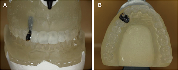
Fig. 1 (A) A frontal view of implant crown that replaced the maxillary right canine, (B) An occlusal view that illustrates the widening of the crown palatal aspect to allow for cross-pin incorporation.

Fig. 2 The 4 fabricated crowns according to the different occlusion schemes. (A) GF, (B) CG, (C) IP, (D) LC.

Fig. 3 The experimental set-up of one crown illustrating the load application and the LDVT arm.
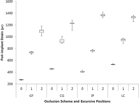
Fig. 4 Box and whisker diagram that outlines the effect of occlusion scheme and excursive position on peri-implant strains.
Cited by 1 articles
-
Load response of the natural tooth and dental implant: A comparative biomechanics study
Dale Robinson, Luis Aguilar, Andrea Gatti, Jaafar Abduo, Peter Vee Sin Lee, David Ackland
J Adv Prosthodont. 2019;11(3):169-178. doi: 10.4047/jap.2019.11.3.169.
Reference
-
1. Kim Y, Oh TJ, Misch CE, Wang HL. Occlusal considerations in implant therapy: clinical guidelines with biomechanical rationale. Clin Oral Implants Res. 2005; 16:26–35.2. Chen YY, Kuan CL, Wang YB. Implant occlusion: biomechanical considerations for implant-supported prostheses. J Dent Sci. 2008; 3:65–74.3. Abichandani SJ, Bhojaraju N, Guttal S, Srilakshmi J. Implant protected occlusion: A comprehensive review. Eur J Prosthodont. 2013; 1:29–36.4. Thornton LJ. Anterior guidance: group function/canine guidance. A literature review. J Prosthet Dent. 1990; 64:479–482.5. Belser UC, Hannam AG. The influence of altered working-side occlusal guidance on masticatory muscles and related jaw movement. J Prosthet Dent. 1985; 53:406–413.6. Okano N, Baba K, Akishige S, Ohyama T. The influence of altered occlusal guidance on condylar displacement. J Oral Rehabil. 2002; 29:1091–1098.7. Ogawa T, Koyano K, Suetsugu T. The relationship between inclination of the occlusal plane and jaw closing path. J Prosthet Dent. 1996; 76:576–580.8. Ogawa T, Koyano K, Suetsugu T. Characteristics of masticatory movement in relation to inclination of occlusal plane. J Oral Rehabil. 1997; 24:652–657.9. The glossary of prosthodontic terms. J Prosthet Dent. 2005; 94:10–92.10. Rinchuse DJ, Kandasamy S, Sciote J. A contemporary and evidence-based view of canine protected occlusion. Am J Orthod Dentofacial Orthop. 2007; 132:90–102.11. Abduo J, Tennant M. Impact of lateral occlusion schemes: A systematic review. J Prosthet Dent. 2015; 114:193–204.12. Abduo J, Tennant M, McGeachie J. Lateral occlusion schemes in natural and minimally restored permanent dentition: a systematic review. J Oral Rehabil. 2013; 40:788–802.13. Carlsson GE. Dental occlusion: modern concepts and their application in implant prosthodontics. Odontology. 2009; 97:8–17.14. Stanford CM, Brand RA. Toward an understanding of implant occlusion and strain adaptive bone modeling and remodeling. J Prosthet Dent. 1999; 81:553–561.15. Weinberg LA. Reduction of implant loading using a modified centric occlusal anatomy. Int J Prosthodont. 1998; 11:55–69.16. Weinberg LA. Therapeutic biomechanics concepts and clinical procedures to reduce implant loading. Part I. J Oral Implantol. 2001; 27:293–301.17. Jemt T, Lindquist L, Hedegard B. Changes in chewing patterns of patients with complete dentures after placement of osseointegrated implants in the mandible. J Prosthet Dent. 1985; 53:578–583.18. Kinsel RP, Lin D. Retrospective analysis of porcelain failures of metal ceramic crowns and fixed partial dentures supported by 729 implants in 152 patients: patient-specific and implant-specific predictors of ceramic failure. J Prosthet Dent. 2009; 101:388–394.19. Rungsiyakull P, Rungsiyakull C, Appleyard R, Li Q, Swain M, Klineberg I. Loading of a single implant in simulated bone. Int J Prosthodont. 2011; 24:140–143.20. Taylor TD, Belser U, Mericske-Stern R. Prosthodontic considerations. Clin Oral Implants Res. 2000; 11:101–107.21. Stanford CM. Issues and considerations in dental implant occlusion: what do we know, and what do we need to find out? J Calif Dent Assoc. 2005; 33:329–336.22. Suit SR, Gibbs CH, Benz ST. Study of gliding tooth contacts during mastication. J Periodontol. 1976; 47:331–334.23. Woda A, Vigneron P, Kay D. Nonfunctional and functional occlusal contacts: a review of the literature. J Prosthet Dent. 1979; 42:335–341.24. Morneburg TR, Pröschel PA. In vivo forces on implants influenced by occlusal scheme and food consistency. Int J Prosthodont. 2003; 16:481–486.
- Full Text Links
-
- Actions
-
Cited
- CITED
-
- Close
- Share
-
- Similar articles
-
- The effects of bone density and crestal cortical bone thickness on micromotion and peri-implant bone strain distribution in an immediately loaded implant: a nonlinear finite element analysis
- Comparative evaluation of peri-implant stress distribution in implant protected occlusion and cuspally loaded occlusion on a 3 unit implant supported fixed partial denture: A 3D finite element analysis study
- Strains around distally inclined implants retaining mandibular overdentures with Locator attachments: an in vitro study
- Occlusal Overload May Affect the Recovery of Peri-implant Bone: A Report of Two Cases
- Blood Vessels of the Peri-Implant Mucosa: a Comparison Between Flap and Flapless Procedures
