Ann Dermatol.
2014 Jun;26(3):411-413.
Neutrophilic Dermatosis Confined to the Lymphedematous Area
- Affiliations
-
- 1Department of Dermatology, Ajou University School of Medicine, Suwon, Korea. maychan@ajou.ac.kr
Abstract
- No abstract available.
MeSH Terms
Figure
-
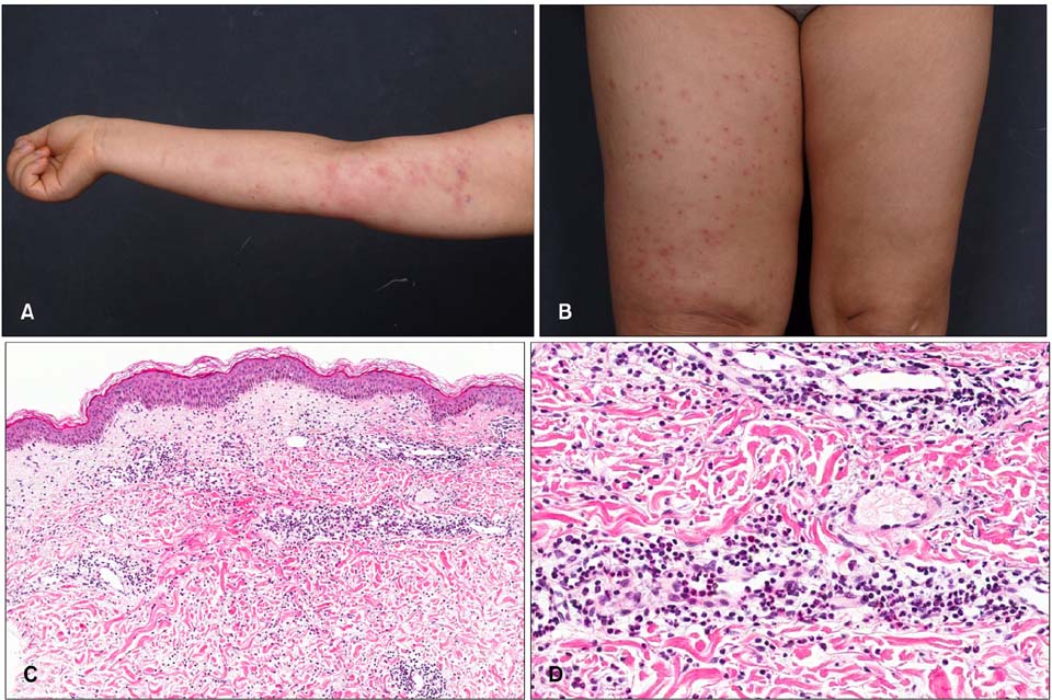
Fig. 1 (A) Case 1. Multiple variable-sized erythematous papules, vesicles, and plaques localized on the right lymphedematous arm. (B) Case 3. Multiple erythematous papules and patches confined on the left lymphedematous leg. (C, D) Histopathological findings. Skin biopsy taken from the patient of case 1, showing marked papillary dermal edema and dense dermal neutrophil infiltrates (H&E; C: ×40, D: ×200).
Reference
-
1. Lee CH, Lee HC, Lu CF, Hsiao CH, Jee SH, Tjiu JW. Neutrophilic dermatosis on postmastectomy lymphoedema: a localized and less severe variant of Sweet syndrome. Eur J Dermatol. 2009; 19:641–642.
Article2. Mallon E, Powell S, Mortimer P, Ryan TJ. Evidence for altered cell-mediated immunity in postmastectomy lymphoedema. Br J Dermatol. 1997; 137:928–933.
Article3. Demitsu T, Tadaki T. Atypical neutrophilic dermatosis on the upper extremity affected by postmastectomy lymphedema: report of 2 cases. Dermatologica. 1991; 183:230–230.
Article4. García-Río I, Pérez-Gala S, Aragüés M, Fernández-Herrera J, Fraga J, García-Díez A. Sweet's syndrome on the area of postmastectomy lymphoedema. J Eur Acad Dermatol Venereol. 2006; 20:401–405.
Article5. Ruocco E, Puca RV, Brunetti G, Schwartz RA, Ruocco V. Lymphedematous areas: privileged sites for tumors, infections, and immune disorders. Int J Dermatol. 2007; 46:662.
Article
- Full Text Links
-
- Actions
-
Cited
- CITED
-
- Close
- Share
-
- Similar articles
-
- Erratum: Neutrophilic Dermatosis Confined to the Lymphedematous Area
- A Case of Neutrophilic Dermatosis of the Hands on Both Palms
- A Case of Acute FEbrile Neutrophilic Dermatosis Following Multiple Keratoacanthoma
- A Case of Steroid-resistant Neutrophilic Dermatosis of the Hands Treated with Dapsone
- A Case of Neutrophilic Dermatosis of the Dorsal Hands with Concomitant Involvement of the Lips
