Spontaneous Absorption of Cerebral Air Embolus Developed Accidentally during an Intra-arterial Procedure
- Affiliations
-
- 1Department of Neurosurgery, School of Medicine, Jeju National University, Jeju, Korea. yfound@hanmail.net
- KMID: 2367332
- DOI: http://doi.org/10.7461/jcen.2016.18.4.391
Abstract
- Cerebral arterial air embolism (CAAE), although infrequent, is a complication that can occur at any time during an invasive medical procedure. We experienced two cases of CAAE during cerebral angiography accidentally. The author reports the two cases of CAAE wherein air emboli dissolved spontaneously and immediately under normal atmospheric pressure, not under therapeutic hyperbaric environment. One of the cases shows entire dissolution of the air embolus on the moving image. This report shows that arterial air embolus can be absorbed spontaneously, and air embolus size is one of the factors that influence air embolus dissolution besides hyperbaric oxygen condition.
Figure
-
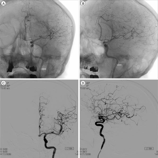
Fig. 1 (A) The captured image of an air embolus (arrow) from the moving image at the distal end of the guiding catheter in the left internal carotid artery. (B) The captured image of an air embolus migrating distally along the ICA (arrow). The anteroposterior (C) and lateral (D) angiograms after the rotational moving image revealed no arterial occlusion in the whole ICA territory. ICA = internal carotid artery.
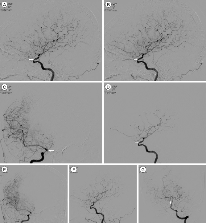
Fig. 2 The angiograms of the right ICA revealed a wandering air embolus (arrow) in the supraclinoid ICA (A, B) and hypoplastic right anterior cerebral artery (C). (D) Angiograms taken three minutes apart showed a decrease in size of the wandering embolus in the right ICA (arrow). The anteroposterior (E) and lateral (F) angiograms of the right ICA demonstrated no air embolus in the entire ICA territory. (G) The left vertebral artery angiogram revealed prominent flow in the posterior communicating artery (arrow). ICA = internal carotid artery.
Cited by 1 articles
-
Spontaneous Absorption of Cerebral Air Emboli
Richard E. Moon
J Cerebrovasc Endovasc Neurosurg. 2017;19(1):52-53. doi: 10.7461/jcen.2017.19.1.52.
Reference
-
1. Annane D, Troche G, Delisle F, Devauchelle P, Hassine D, Paraire F, et al. Kinetics of elimination and acute consequences of cerebral air embolism. J Neuroimaging. 1995; 7. 5(3):183–189. PMID: 7626827.
Article2. Bauerle J, Fischer A, Hornig T, Egger K, Wengenmayer T, Bardutzky J. Therapeutic hypothermia in cerebral air embolism: a case report. Springerplus. 2013; 8. 2:411. PMID: 24024097.
Article3. Benson J, Adkinson C, Collier R. Hyperbaric oxygen therapy of iatrogenic cerebral arterial gas embolism. Undersea Hyperb Med. 2003; Summer. 30(2):117–126. PMID: 12964855.4. Dexter F, Hindman BJ. Recommendations for hyperbaric oxygen therapy of cerebral air embolism based on a mathematical model of bubble absorption. Anesth Analg. 1997; 6. 84(6):1203–1207. PMID: 9174293.
Article5. Fritz H, Hossmann KA. Arterial air embolism in the cat brain. Stroke. 1979; Sep-Oct. 10(5):581–589. PMID: 41347.
Article6. Gupta R, Vora N, Thomas A, Crammond D, Roth R, Jovin T, et al. Symptomatic cerebral air embolism during neuro-angiographic procedures: incidence and problem avoidance. Neurocrit Care. 2007; 7(3):241–246. PMID: 17805494.
Article7. McDermott JJ, Dutka AJ, Koller WA, Pearson RR, Flynn ET. Comparison of two recompression profiles in treating experimental cerebral air embolism. Undersea Biomed Res. 1992; 5. 19(3):171–185. PMID: 1595138.9. Reasoner DK, Dexter F, Hindman BJ, Subieta A, Todd MM. Somatosensory evoked potentials correlate with neurological outcome in rabbits undergoing cerebral air embolism. Stroke. 1996; 10. 27(10):1859–1864. PMID: 8841345.
Article10. Sayama T, Mitani M, Inamura T, Yagi H, Fukui M. Normal diffusion-weighted imaging in cerebral air embolism complicating angiography. Neuroradiology. 2000; 3. 42(3):192–194. PMID: 10772140.
Article11. Surve RM, Reddy KR, Bansal S, Ramalingaiah A. Massive cerebral air embolism during stent-assisted coiling of internal carotid artery aneurysm. Neurol India. 2013; Jan-Feb. 61(1):95–97. PMID: 23466861.
Article12. Tan LA, Keigher KM, Lopes DK. Symptomatic cerebral air embolism during stent-assisted coiling of an unruptured middle cerebral artery aneurysm: intraoperative diagnosis and management of a rare complication. J Cerebrovasc Endovasc Neurosurg. 2014; 6. 16(2):93–97. PMID: 25045648.
Article13. Tsetsou S, Eeckhout E, Qanadli SD, Lachenal Y, Vingerhoets F, Michel P. Nonaccidental arterial cerebral air embolism: a ten-year stroke center experience. Cerebrovasc Dis. 2013; 35(4):392–395. PMID: 23635471.
Article14. Voorhies RM, Fraser RA. Cerebral air embolism occurring at angiography and diagnosed by computerized tomography. Case report. J Neurosurg. 1984; 1. 60(1):177–178. PMID: 6689713.15. Yesilaras M, Atilla OD, Aksay E, Kilic TY. Retrograde cerebral air embolism. Am J Emerg Med. 2014; 12. 32(12):1562.e1–1562.e2.
Article
- Full Text Links
-
- Actions
-
Cited
- CITED
-
- Close
- Share
-
- Similar articles
-
- Spontaneous Absorption of Cerebral Air Emboli
- Captured Macro-embolus of Fractured Atheromatous Plaque by the Embolic Protection Device during Carotid Stent Assisted Angioplasty
- Cerebral Air Embolism Following a Gastroscopy
- Symptomatic Cerebral Air Embolism During Stent-assisted Coiling of an Unruptured Middle Cerebral Artery Aneurysm: Intraoperative Diagnosis and Management of a Rare Complication
- Successful Intra-arterial Stent Thrombectomy in Acute Infarction Caused by Spontaneous Middle Cerebral Artery Dissection
