A Gene and Neural Stem Cell Therapy Platform Based on Neuronal Cell Type-Inducible Gene Overexpression
- Affiliations
-
- 1Department of Neurosurgery, Spine & Spinal Cord Institute and Brain Korea 21 PLUS Project for Medical Science, Yonsei University College of Medicine, Seoul, Korea. hayoon@yuhs.ac
- 2Department of Bioengineering, College of Engineering, Hanyang University, Seoul, Korea.
- KMID: 2366346
- DOI: http://doi.org/10.3349/ymj.2015.56.4.1036
Abstract
- PURPOSE
Spinal cord injury (SCI) is associated with permanent neurological damage, and treatment thereof with a single modality often does not provide sufficient therapeutic outcomes. Therefore, a strategy that combines two or more techniques might show better therapeutic effects.
MATERIALS AND METHODS
In this study, we designed a combined treatment strategy based on neural stem cells (NSCs) introduced via a neuronal cell type-inducible transgene expression system (NSE::) controlled by a neuron-specific enolase (NSE) promoter to maximize therapeutic efficiency and neuronal differentiation. The luciferase gene was chosen to confirm whether this combined system was working properly prior to using a therapeutic gene. The luciferase expression levels of NSCs introduced via the neuronal cell type-inducible luciferase expression system (NSE::Luci) or via a general luciferase expressing system (SV::Luci) were measured and compared in vitro and in vivo.
RESULTS
NSCs introduced via the neuronal cell type-inducible luciferase expressing system (NSE::Luci-NSCs) showed a high level of luciferase expression, compared to NSCs introduced via a general luciferase expressing system (SV::Luci-NSCs). Interestingly, the luciferase expression level of NSE::Luci-NSCs increased greatly after differentiation into neurons.
CONCLUSION
We demonstrated that a neuronal cell type-inducible gene expression system is suitable for introducing NSCs in combined treatment strategies. We suggest that the proposed strategy may be a promising tool for the treatment of neurodegenerative disorders, including SCI.
Keyword
MeSH Terms
-
Cell Differentiation/genetics/physiology
*Gene Expression
Gene Regulatory Networks
*Genetic Therapy
Humans
Luciferases/genetics/*metabolism
*Neural Stem Cells
Neurons/metabolism
Phosphopyruvate Hydratase/metabolism
Promoter Regions, Genetic
Spinal Cord Injuries/*therapy
Stem Cells/*metabolism
Luciferases
Phosphopyruvate Hydratase
Figure
-

Fig. 1 Plasmid construction. Plasmid construction of neuronal cell type-inducible (NSE::Luci) or general (SV::Luci) luciferase overexpression vector. The neuron-specific enolase (NSE) promoter region was inserted to pGL3-basic vector after digestion (NheI and BglII sites).
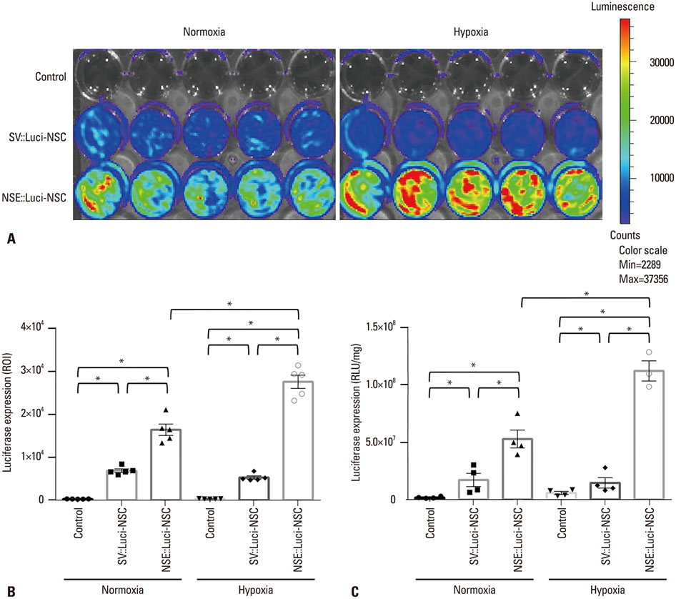
Fig. 2 Luciferase expression pattern of neural stem cells introduced with a neuronal cell type-inducible luciferase overexpression system (NSE::Luci-NSCs) in normoxia and hypoxia. Luciferase expression levels of Control, SV::Luci-NSCs, and NSE::Luci-NSCs examined by luciferase expression imaging at 24-48 h after transfection. (A) The result of luciferase expression imaging indicates that luciferase in NSE::Luci-NSCs group is highly over-expressed, compared to the other groups. The color scale bar indicates the luciferase expression level. (B) Quantitative analysis of luciferase expression imaging in (A). (C) Luciferase expression levels of Control, SV::Luci-NSCs, and NSE::Luci-NSCs examined by luciferase assay at 24-48 h after transfection. Luciferase expression of NSE::Luci-NSC is much higher than the other groups in normoxia and hypoxia. The number of the symbol indicates the value of n. Data are represented as mean±standard error of the mean. *p<0.05. ROI, regions of interest; NSC, neural stem cell; NSE, neuron-specific enolase; RLU, relative light units.

Fig. 3 Neuronal cell-type-inducible luciferase gene overexpression systems in non-neuronal cells. Luciferase expression levels of SV::Luci-293FT and NSE::Luci-293FT confirmed by luciferase assay at 48 h transfection. The number of the symbol indicates the value of n. Data are represented as mean±standard error of the mean. *p<0.05. NSE, neuron-specific enolase; RLU, relative light units.
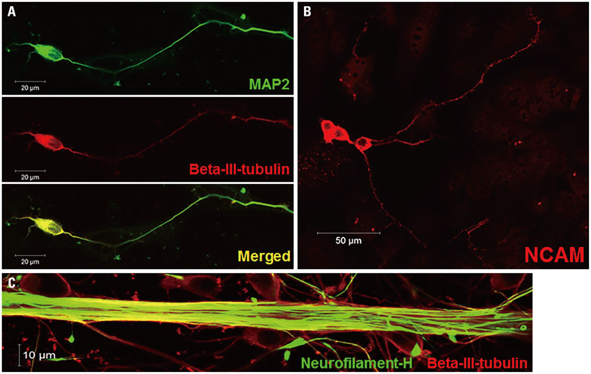
Fig. 4 Neuronal differentiation potency. Neuronal differentiation potency of NSCs was confirmed by fluorescence staining specific for each neuron. (A and B) Most of the differentiated cells are positive for neuron-specific markers, such as beta-III-tubulin, MAP2, and NCAM. (C) The thick axonal bundle was confirmed by staining with neurofilament and beta-III-tubulin. NSC, neural stem cell; MAP2, microtubule-associated protein 2; NCAM, neural cell adhesion molecule.
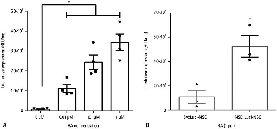
Fig. 5 High luciferase expression pattern after differentiation into neurons. (A) The luciferase expression level in neural stem cells and differentiated neurons was compared by luciferase assay. Luciferase expression is significantly greater in differentiated neurons (with RA) than in neural stem cells (without RA). (B) The luciferase expression level of NSE::Luci-NSCs after neuronal differentiation is significantly greater than that of SV::Luci-NSCs after differentiation. The number of the symbol indicates the value of n. Data are represented as mean±standard error of the mean. *p<0.05. RA, retinoic acid; NSC, neural stem cell; NSE, neuron-specific enolase; RLU, relative light units.
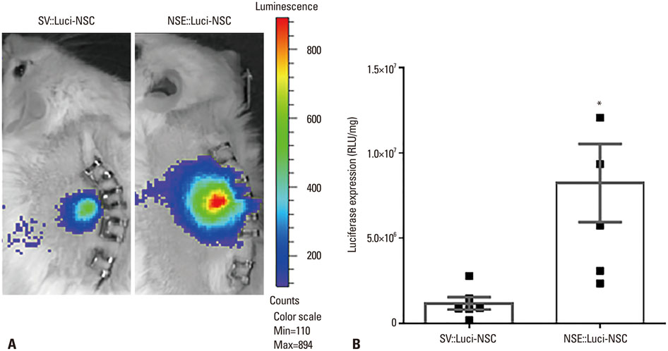
Fig. 6 Luciferase expression pattern of neuronal cell-type-inducible luciferase gene overexpressing neural stem cells after transplantation. (A) Luciferase expression pattern of SV::Luci-NSCs and NSE::Luci-NSCs confirmed by IVIS 24-48 h after cell transplantation. NSE::Luci-NSCs transplanted into the spinal cord show a high luciferase expression, compared to SV::Luci-NSCs, after transplantation. The color scale indicates the luciferase expression level, not the cell numbers in this study. (B) Luciferase expression pattern of SV::Luci-NSCs and NSE::Luci-NSCs confirmed by luciferase assay 24-48 h after cell transplantation. NSE::Luci-NSCs transplanted into the spinal cord consistently show a high luciferase expression pattern, compared to SV::Luci-NSCs, after transplantation. The number of the symbol indicates the value of n. Data are represented as mean±standard error of the mean. *p<0.05. NSE, neuron-specific enolase; NSC, neural stem cell; RLU, relative light units; IVIS, In Vivo Imaging System.
Reference
-
1. Hagg T, Oudega M. Degenerative and spontaneous regenerative processes after spinal cord injury. J Neurotrauma. 2006; 23:264–280.
Article2. Lee M, Lee ES, Kim YS, Choi BH, Park SR, Park HS, et al. Ischemic injury-specific gene expression in the rat spinal cord injury model using hypoxia-inducible system. Spine (Phila Pa 1976). 2005; 30:2729–2734.
Article3. Jin H, Liu ML, Kim HA, Lee M, An S, Oh J, et al. Role of the oxygen-dependent degradation domain in a hypoxia-inducible gene expression system in vascular endothelial growth factor gene therapy. Spine (Phila Pa 1976). 2009; 34:E952–E958.
Article4. Navarro V, Millecamps S, Geoffroy MC, Robert JJ, Valin A, Mallet J, et al. Efficient gene transfer and long-term expression in neurons using a recombinant adenovirus with a neuron-specific promoter. Gene Ther. 1999; 6:1884–1892.
Article5. Tsuchiya R, Yoshiki F, Kudo Y, Morita M. Cell type-selective expression of green fluorescent protein and the calcium indicating protein, yellow cameleon, in rat cortical primary cultures. Brain Res. 2002; 956:221–229.
Article6. Kügler S, Kilic E, Bähr M. Human synapsin 1 gene promoter confers highly neuron-specific long-term transgene expression from an adenoviral vector in the adult rat brain depending on the transduced area. Gene Ther. 2003; 10:337–347.
Article7. Iwai H, Nori S, Nishimura S, Yasuda A, Takano M, Tsuji O, et al. Transplantation of neural stem/progenitor cells at different locations in mice with spinal cord injury. Cell Transplant. 2014; 23:1451–1464.
Article8. Lu P, Wang Y, Graham L, McHale K, Gao M, Wu D, et al. Long-distance growth and connectivity of neural stem cells after severe spinal cord injury. Cell. 2012; 150:1264–1273.
Article9. Liu ML, Oh JS, An SS, Pennant WA, Kim HJ, Gwak SJ, et al. Controlled nonviral gene delivery and expression using stable neural stem cell line transfected with a hypoxia-inducible gene expression system. J Gene Med. 2010; 12:990–1001.
Article10. Kim HM, Hwang DH, Lee JE, Kim SU, Kim BG. Ex vivo VEGF delivery by neural stem cells enhances proliferation of glial progenitors, angiogenesis, and tissue sparing after spinal cord injury. PLoS One. 2009; 4:e4987.
Article11. Kim HJ, Oh JS, An SS, Pennant WA, Gwak SJ, Kim AN, et al. Hypoxia-specific GM-CSF-overexpressing neural stem cells improve graft survival and functional recovery in spinal cord injury. Gene Ther. 2012; 19:513–521.
Article12. Oh JS, An SS, Gwak SJ, Pennant WA, Kim KN, Yoon DH, et al. Hypoxia-specific VEGF-expressing neural stem cells in spinal cord injury model. Neuroreport. 2012; 23:174–178.
Article13. An SS, Jin HL, Kim KN, Kim DS, Cho J, Liu ML, et al. Neuroprotective effect of combined hypoxia-induced VEGF and bone marrow-derived mesenchymal stem cell treatment. Childs Nerv Syst. 2010; 26:323–331.
Article14. Lu P, Jones LL, Snyder EY, Tuszynski MH. Neural stem cells constitutively secrete neurotrophic factors and promote extensive host axonal growth after spinal cord injury. Exp Neurol. 2003; 181:115–129.
Article
