Treatment of Fused Knee Flexion and Valgus Deformity Using Gradual Correction with Ilizarov Frame
- Affiliations
-
- 1Department of Orthopedic Surgery, Yeungnam University Medical Center, Daegu, Korea. ossoj@med.yu.ac.kr
- KMID: 2364306
- DOI: http://doi.org/10.4055/jkoa.2016.51.6.527
Abstract
- Various surgical methods including soft tissue procedures and bone procedures are commonly used to treat knee flexion and contracture deformity. However, several complications such as, neurovascular injury and skin necrosis were reported because of rapid correction. We aim to report good results from gradual correction using Ilizarov following supracondylar osteotomy in a 24-year-old man suffering from fused knee flexion and valgus deformity, a complication developed by septic arthritis.
MeSH Terms
Figure
-

Figure 1 Preoperative photographs show flexion and valgus deformity of right knee.

Figure 2 Preoperative anteroposterior (A) and lateral (B) radiographs show a right fused knee joint with valgus and flexion deformity.
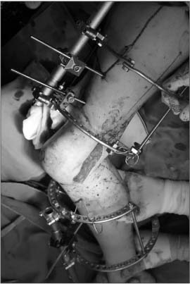
Figure 3 Intraoperative photograph shows correcting flexion and valgus deformity with Ilizarov following supracondylar osteotomy through lateral approach.
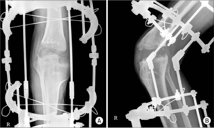
Figure 4 Immediate postoperative anteroposterior (A) and lateral (B) radiographs show a knee joint, corrected to preoperatively planed valgus and flexion angle with Ilizarov.

Figure 5 Second postoperative anteroposterior (A) and lateral (B) radiographs show a corrected knee joint which is fixated using pre-bended locking plate.

Figure 6 Radiographs taken at 3 months after second surgery show union of knee joint with corrected flexion and valgus deformity.
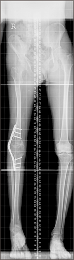
Figure 7 Long standing anteroposterior radiograph taken at 3 months after surgery shows leg length discrepancy of right lower leg.
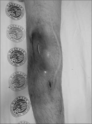
Figure 8 Photographs taken at 3 years after surgery show an exposed plate with open wound.
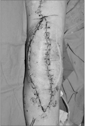
Figure 9 Intaoperative photograph shows wound coverage using reverse anterolateral thigh island flap.
Reference
-
1. Klatt J, Stevens PM. Guided growth for fixed knee flexion deformity. J Pediatr Orthop. 2008; 28:626–631.
Article2. Damsin JP, Ghanem I. Treatment of severe flexion deformity of the knee in children and adolescents using the Ilizarov technique. J Bone Joint Surg Br. 1996; 78:140–144.
Article3. Hosny GA, Fadel M. Managing flexion knee deformity using a circular frame. Clin Orthop Relat Res. 2008; 466:2995–3002.
Article4. Gaurav K, Vilas J. A new approach to the management of fixed flexion deformity of the knee using Ilizarov's principle of distraction histogenesis: a preliminary communication. Int J Low Extrem Wounds. 2010; 9:70–73.
Article5. Gartsman GM, Bennett JB, Cain TE. Surgical correction of severe knee pterygium. Microsurgery. 1988; 9:246–248.
Article6. Paley D. Problems, obstacles, and complications of limb lengthening by the Ilizarov technique. Clin Orthop Relat Res. 1990; (250):81–104.
Article7. Tomak Y, Piskin A, Gulman B, Tomak L. Treatment of U-shaped bone ankylosis of the knee with the Ilizarov method. A case report. J Bone Joint Surg Am. 2005; 87:1104–1107.8. Zouari O, Gargouri A, Jenzri M, Hadinane R, Slimane N. Supracondylar femoral extension osteotomy for knee flexion contracture correction in poliomyelitic conditions. Rev Chir Orthop Reparatrice Appar Mot. 2001; 87:361–366.9. Jeong BO, Kim TY, Song WJ. Use of Ilizarov external fixation without soft tissue release to correct severe, rigid equinus deformity. J Foot Ankle Surg. 2015; 54:821–825.
Article10. Weiner DS, Tank JC, Jonah D, et al. An operative approach to address severe genu valgum deformity in the Ellis-van Creveld syndrome. J Child Orthop. 2014; 8:61–69.
Article
- Full Text Links
-
- Actions
-
Cited
- CITED
-
- Close
- Share
-
- Similar articles
-
- Ilizarov Correction for Knee Flexion Contracture
- Treatment of genu recurvatum with the Ilizarov external fixator and proximal tibial corticotomy
- Supracondylar Extension osteotomy for Knee Flexion Contracture by the Ilizarov Technique
- Surgical Treatment for the Knee Flexion Deformity in Spastic Cerebral Palsy
- Total Knee Arthroplasty for Severe Flexion Contracture in Rheumatoid Arthritis Knees
