Sinonasal sarcoidosis of the maxillary sinus and infraorbital nerve: a case report
- Affiliations
-
- 1Department of Oral and Maxillofacial Surgery, Kunhitharuvai Memorial Charitable Trust (KMCT) Dental College and Hospitals, Kozhikode, India. shermil12@gmail.com
- 2Department of Oral Medicine and Radiology, Kunhitharuvai Memorial Charitable Trust (KMCT) Dental College and Hospitals, Kozhikode, India.
- KMID: 2364021
- DOI: http://doi.org/10.5125/jkaoms.2015.41.4.217
Abstract
- Sinonasal sarcoidosis in the head and neck region is infrequent. Its occurrence can be either isolated in combination with other systems. The literature reveals that the occurrence of sinonasal sarcoidosis without lung involvement is rare. In general, sarcoidosis is a chronic non-caseating granulomatous disease of unknown origin, often identified after biopsy. In this article, we report on a benign tumor of the face that produced a diagnostic dilemma, necessitating refinement of the surgical access and in toto removal of the benign tumor.
Figure
-
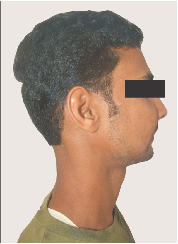
Fig. 1 Preoperative swelling on the right midface region.
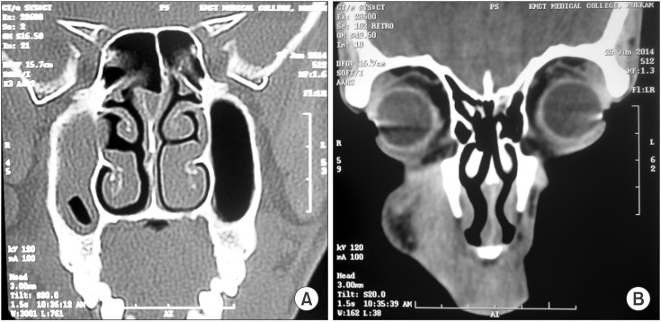
Fig. 2 Computed tomographies show sinus lesion (A) and soft tissue lesion (B).
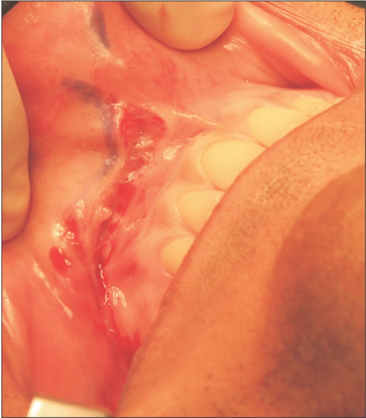
Fig. 3 Placement of intraoral vestibular incision to gain access to the lesion.
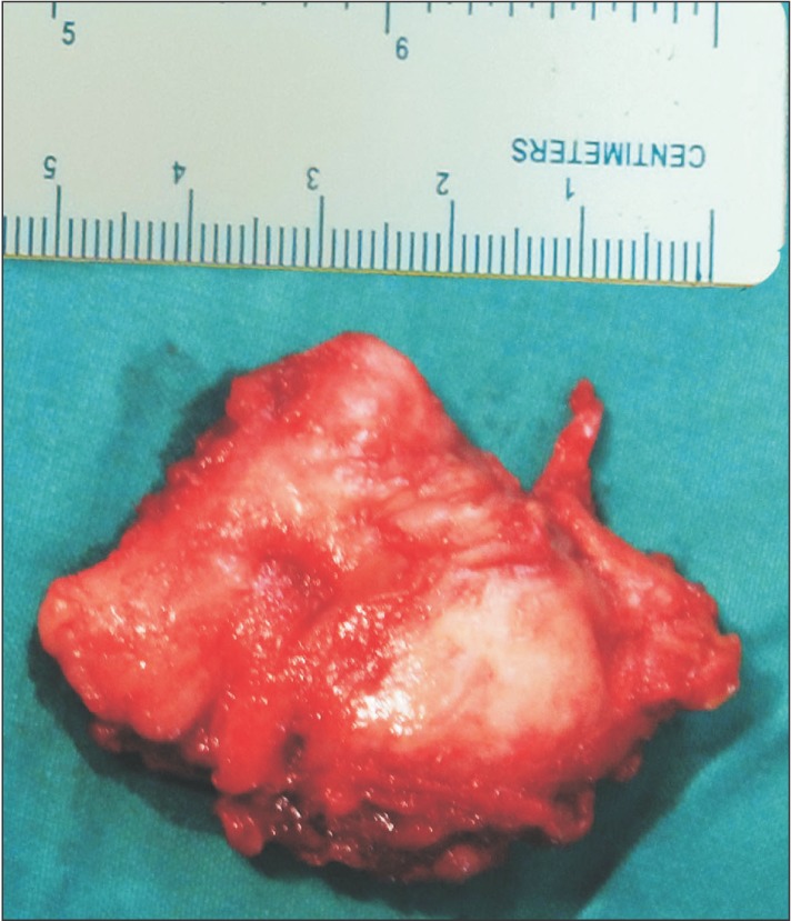
Fig. 4 Excised mass in toto.
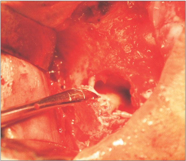
Fig. 5 Nerve relation with the tumor.
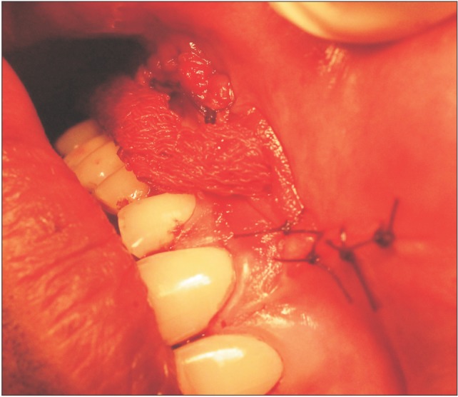
Fig. 6 Antral pack and closure.
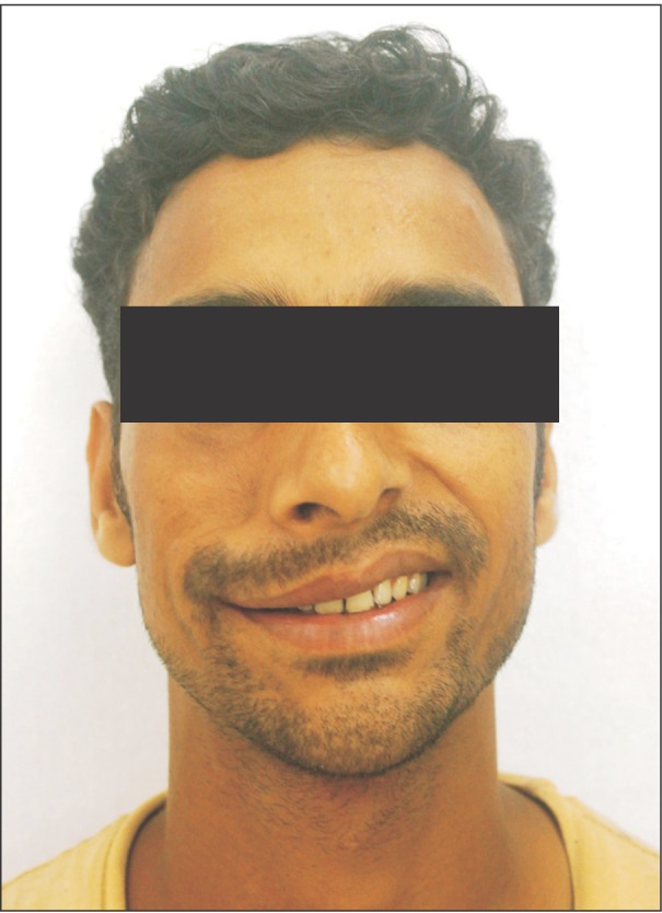
Fig. 7 One-month postoperative.
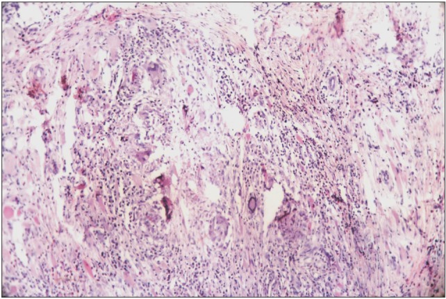
Fig. 8 H&E stained low power (×10) section shows abundant chronic inflammatory cells interspersed with different type of multinucleated giant cells.
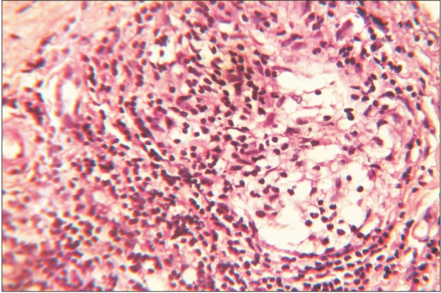
Fig. 9 H&E stained high power (×40) section shows chronic inflammatory cells.
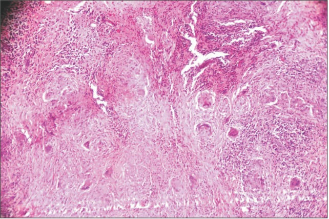
Fig. 10 H&E stained low power (×10) section shows chronic inflammatory cells, multinucleated giant cells and connective tissue.
Cited by 1 articles
-
Middle superior and anterior superior alveolar nerve injury following trauma to the maxillary sinus: a prospective clinico-radiographic evaluation
Sathish Radhakrishna, Eashwari Narayanan
J Korean Assoc Oral Maxillofac Surg. 2023;49(5):262-269. doi: 10.5125/jkaoms.2023.49.5.262.
Reference
-
1. Scadding JG. Sarcoidosis. London: Eyre & Spottiswoode;1967.2. Braun JJ, Gentine A, Pauli G. Sinonasal sarcoidosis: review and report of fifteen cases. Laryngoscope. 2004; 114:1960–1963. PMID: 15510022.
Article3. Dessouky OY. Isolated sinonasal sarcoidosis with intracranial extension: case report. Acta Otorhinolaryngol Ital. 2008; 28:306–308. PMID: 19205596.4. Mallis A, Mastronikolis NS, Koumoundourou D, Stathas T, Papadas TA. Sinonasal sarcoidosis: a case report. Eur Rev Med Pharmacol Sci. 2010; 14:1097–1099. PMID: 21375142.5. deShazo RD, O'Brien MM, Justice WK, Pitcock J. Diagnostic criteria for sarcoidosis of the sinuses. J Allergy Clin Immunol. 1999; 103:789–795. PMID: 10329811.
Article6. Neville E, Mills RG, Jash DK, Mackinnon DM, Carstairs LS, James DG. Sarcoidosis of the upper respiratory tract and its association with lupus pernio. Thorax. 1976; 31:660–664. PMID: 1013937.
Article7. Crystal RG. Sarcoidosis. In : Wonsiewicz M, Englis MR, editors. Harrison's principles of internal medicine. 16th ed. New York: McGraw-Hill;2005. p. 2022–2023.8. Krespi YP, Kuriloff DB, Aner M. Sarcoidosis of the sinonasal tract: a new staging system. Otolaryngol Head Neck Surg. 1995; 112:221–227. PMID: 7838542.
Article9. Long CM, Smith TL, Loehrl TA, Komorowski RA, Toohill RJ. Sinonasal disease in patients with sarcoidosis. Am J Rhinol. 2001; 15:211–215. PMID: 11453511.
Article10. Kirsten AM, Watz H, Kirsten D. Sarcoidosis with involvement of the paranasal sinuses: a retrospective analysis of 12 biopsy-proven cases. BMC Pulm Med. 2013; 13:59. PMID: 24070015.
Article11. Mazziotti S, Gaeta M, Blandino A, Vinci S, Pandolfo I. Perineural spread in a case of sinonasal sarcoidosis: case report. AJNR Am J Neuroradiol. 2001; 22:1207–1208. PMID: 11415921.
